Can CT pinpoint the cause of acute chest pain in the emergency room? The idea may raise eyebrows, but let's face it: CT is already being used in the ED to evaluate everything but the scrub sink. And for quickly assessing the nebulous and sundry causes of chest pain, it's not as though doctors have a lot of alternatives.
So researchers from the University of Maryland School of Medicine in Baltimore decided to give it a go -- scanning 69 clinically stable patients who presented to the emergency department with acute chest pain. They examined the data for coronary calcium and stenosis, ejection fraction, wall motion and perfusion abnormalities, and significant noncardiac causes of chest pain, such as pulmonary embolism, dissection, and pneumonia.
CT was both sensitive and specific, as it turned out.
"Substantial advances have occurred in patient evaluation and triage over the past decade, but the assessment of chest pain in the emergency department remains a significant challenge," wrote Drs. Charles White, Dick Kuo, Mark Kelemen, and colleagues in the American Journal of Roentgenology (AJR, August 2005, Vol. 185, pp. 533-540).
Of course, cardiac or noncardiac diagnoses may be immediately apparent in some patients, but in others the initial clinical evaluation is often equivocal, resulting in a high proportion of hospital admissions, they wrote.
"Nevertheless, it is estimated that 4%-8% of patients are inappropriately discharged from the emergency department and ultimately prove to have a myocardial infarction, the most important cause of acute chest pain," the team wrote.
In addition, the clinical pathway for diagnosing acute cardiac ischemia, including acute myocardial infarction and unstable angina, is guided by patient history, risk factors, and ECG results, a process that is known to lack sensitivity. Serum markers can be helpful, but most levels rise too slowly to be helpful for triage.
For their prospective study, the researchers recruited patients who presented in the emergency department with acute chest pain between November 2003 and July 2004, and classified them using a scale of 1-5 based on initial clinical impression. Category 1 patients had acute myocardial infarction and were excluded as clinically unstable. Category 2 patients were considered to have definite angina with uncertainty regarding acute myocardial infarction. Patients classified as 3 had probable angina, and those at 4 probably did not have angina, but still had suspicion of significant noncoronary chest pain. Category 5 patients were considered unlikely to have a significant cause of chest pain and were also excluded. Ultimately, 69 patients participated in the study, the authors wrote.
"The emergency department physician was asked in each case before the study whether CT would have been performed for the conventional workup," the team wrote. "The CT study was done early in the clinical evaluation of the patient in the interval immediately after the ECG was done and blood samples drawn and before a decision was made as to further care or studies for the patient and before the results of cardiac enzymes were available."
IV beta blockers were used to control heart rates higher than 70 beats per minute (bpm). Retrospective ECG-gated CT images (75% of the RR interval) were obtained on a 16-slice MX 800IDT scanner (Philips Medical Systems, Andover, MA) during a single breath-hold, though patients were advised to exhale slowly if they couldn't hold their breath throughout the 30-second scan.
Following administration of 120-150 mL of iodinated contrast material at 3-4 mL/sec, patients were scanned using 140 kVp, 350-500 mAs, pitch 0.2-0.3, and rotation time of 0.42 seconds. Automated bolus timing was performed using a threshold of 150 HU, with the region of interest over the aorta.
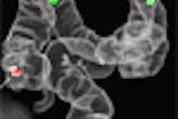
"Specific noncardiac entities that were evaluated included, but were not limited to, aortic dissection, pulmonary embolism, pneumonia, pneumothorax, pericardial effusion, and rib fracture," the team wrote. "A qualitative assessment of the presence of coronary artery calcifications was made."
The ejection fraction was calculated for each dataset, and curved planar reconstructions were generated for each of the coronary arteries. Wall motion was assessed qualitatively on a cine loop for areas of hypokinesis or akinesis, the authors wrote.
Of the 69 patients, 45 (65%) wouldn't have otherwise had a CT scan, the team reported. There were no significant findings in nearly three-fourths of the patients (52/69), who were diagnosed with clinically insignificant chest pain, the authors wrote.
"Thirteen patients (19%) had significant CT findings concordant with the final diagnosis," the team wrote. These included 10 patients with positive CT diagnosis of angina due to cardiac disease, and three patients had pericarditis with moderately large pericardial effusion.
"CT failed to suggest a diagnosis in two patients (3%), both of whom proved to have clinically significant coronary artery stenoses," the authors wrote. The two stenoses were identified on coronary angiography, and were located in the left anterior descending and first diagonal branches, respectively.
Overall, CT was 83% sensitive and 96% specific for establishing a cardiac cause of chest pain. For noncardiac chest pain, CT was 87% sensitive and 96% specific.
At follow-up one to two months later, final diagnoses were made by consensus by an emergency department physician, a cardiologist, and a radiologist, based on evaluation of all available data.
"ECG-gated MDCT appears to be logistically feasible, and shows promise as a comprehensive method for evaluating cardiac and noncardiac chest pain in stable emergency department patients," the authors concluded. "Further hardware and software improvements will be necessary for the adoption of this paradigm in clinical practice."
CT is effective in diagnosing many conditions that may not be apparent on initial clinical evaluation, including pneumonia, aortic dissection, pulmonary embolism, and coronary stenosis, they noted.
Still, the group cautioned that diagnosing the cause of acute chest pain "remains a formidable task because of extensive etiology that ranges from benign to potentially lethal. The evaluation of many of these conditions, particularly the conventional assessment for the presence of cardiac disease in the acute setting, is often inconclusive and may require further invasive testing."
By Eric Barnes
AuntMinnie.com staff writer
August 15, 2005
Related Reading
Chest pain widespread but not tied to coronary calcium, July 20, 2005
Canadian cardiologists define low risk for women with chest pain, May 30, 2005
Chest pain warrants closer scrutiny in some patients, November 29, 2004
Definitive diagnostic testing useful in ER patients with non-MI chest pain , July 21, 2003
Cardiac MRI may improve triage of patients with chest pain, February 4, 2003
Copyright © 2005 AuntMinnie.com