SAN FRANCISCO - Electron-beam CT is undergoing a renaissance of sorts as new scanner technology hits the market. At the same time, many facilities are starting to get a better feel for the performance of the new 16-detector-row multislice spiral CT scanners in CT angiography (CTA) applications. The confluence of new system introductions makes it an ideal time to revisit the question of which scanner gets to the heart of accuracy.
According to Dr. Friedrich Knollmann from the Klinik für Strahlenheilkunde at Berlin's Charité Hospital, CTA results have improved significantly in both scanner types, although there is still room for improvement. According to his group's research, and that of other investigators, it is the application that determines which scanner will produce the best results in cardiac applications.
"Cardiac CT is a little bit like landing in San Francisco," Knollmann said. "It is certainly an enjoyable view, although occasionally there is a bit of mist that demands a closer look." Knollmann discussed his evaluation methods, especially in terms of temporal and spatial resolution of the two scanner types, the clinical implications in the literature, and prospects for future developments, on Saturday at the 2003 Symposium on Multidetector-Row CT, sponsored by Stanford University of Stanford, CA.
Resolution in action
Spatial resolution is one important measure of performance. When optimized, it permits the evaluation of smaller branch arteries, and increases diagnostic confidence. Here, multislice CT retains its edge, Knollmann said. For example, the new e-Speed EBCT scanner from GE Medical Systems of Waukesha, WI, has spatial resolution of 1.5 mm, while submillimeter resolution is available on the 16-row multislice machines.
In terms of temporal resolution, EBCT beats MDCT by default, Knollmann said, adding that higher temporal resolution can't compensate for low spatial resolution. "If we use our current system, the C-150 scanner for coronary angiography (GE Medical Systems), we are limited to 3-mm slice thicknesses, and although we will routinely achieve 100 milliseconds of temporal resolution, we are in a worse position to evaluate all the coronary artery segments," Knollmann said.
In contrast, a 16-row MDCT scanner has a default temporal resolution of 250 msec at 0.5-second gantry rotation time, which can be improved even further with the aid of 0.4-second rotation speed, and special segmented reconstruction algorithms offered by some MDCT vendors. However, the use of such algorithms doesn't necessarily improve spiral CT's temporal-resolution shortcomings in clinical practice.
"Even if we use a two-segment reconstruction algorithm with retrospectively gated MDCT, it may not (improve temporal resolution) in all instances because of the prerequisite of a perfectly stable heart rate," Knollmann said, noting that further developments are afoot in terms of new algorithms that may be more clinically robust than the current solutions.
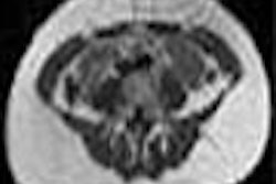
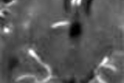
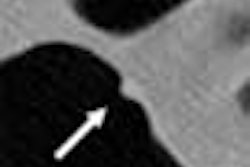
In the moving heart, dynamic spatial resolution is also key, he said. So in order to determine which scanner performs better in this area, Knollmann and colleagues created a phantom that uses a moving tungsten wire that creates a 0.50-micron point of light on CT. The group used this phantom to evaluate image quality in both acquisition methods.
Using 3-mm slice thickness (FOV 90 mm, 512 matrix, 100-ms scan), EBCT (GE C-150) produced temporal resolution as low as 150 msec, and in-plane spatial resolution of 7.3 to 7.8 line pairs per cm. The LightSpeed 16 multidetector-row scanner (GE Medical Systems) yielded temporal resolution of 250 msec, with higher in-plane spatial resolution compared to EBCT.
The new multislice mode in EBCT scanners is not useful in that it doesn't improve spatial resolution enough to evaluate coronary segments, he added. (Knollman acknowledged that while his group cited manufacturer's data for comparison purposes, it has not installed GE's latest EBCT scanner, which the company calls its first completely new EBCT system since it acquired EBCT developer Imatron in 2001.)
A point-spread formula was used to gauge the variations that occurred while imaging the point of light in a given location. The measured variations were much higher, and therefore inferior, in MDCT, though retrospective gating helps to compensate for it.
"In clinical practice using a retrospective ECG-gated method... we can select the best time point during the cardiac cycle that we would use for diagnosis," Knollmann said. This method creates additional datasets that can be used when needed to improve diagnostic confidence. Of course, retrospective gating also necessitates a higher radiation dose than EBCT's prospective gating.
Testing the traditional retrospective multislice reconstruction method, the group created volume renderings covering from 0% to 90% of the RR interval; only at 60%, 70%, and 80% of the RR interval were the right coronary arteries visible at all, Knollmann said. The group then picked the best rendering of each vessel for comparison with EBCT.
Retrospective gating is not available in EBCT, but something approaching its effect is now possible in so-called multiphasic acquisition, which allows for up to three acquisitions per cardiac cycle in the same slice position, Knollmann said.
In order to determine the best visual data quality acquired from each slice position over the device cycle, Knollmann and colleagues compared the selected MDCT reconstructions with the best EBCT images acquired at 40% and 60% of the RR interval. They measured the performance of the LightSpeed 16 16-slice MDCT and the C-150 EBCT scanners at four different heart rates.
The results, based on the segmented reconstruction method, showed lower in-plane spatial resolution in the multiphasic acquisitions of the EBCT scanner across the entire resolution parameter. "The amazing thing was that at higher heart rates, the spatial resolution in plane decreased significantly with electron beam, though it didn't with the helical method," Knollmann said. "In a dynamic situation, the spatial resolution is apparently much better with the helical system."
Clinical considerations
According to Knollmann, important clinical perspective on the performance of the two scanner types is provided in three papers by Dr. Stephan Achenbach and colleagues from the University of Erlangen-Nürnberg, Germany (New England Journal of Medicine, December 1998, Vol. 339: 27, pp. 1964-1971; Circulation 2001, Vol. 103, pp. 2535-2538; Circulation 2003, Vol. 107, pp. 664-666).
Following the article order above, sensitivity for the detection of coronary stenoses was 92%, 91%, and 85% for EBCT, four-row MDCT, and 12-row MDCT, respectively. Specificity was 94%, 84%, and 78%, respectively. The rate of evaluable cases was 61%, 30%, and 74%, respectively.
"With EBCT there were 61% of patients in whom all (proximal) segments could be evaluated," Knollmann said. But, he added, the fact that evaluable segments dropped to 30% for the four-row MDCT system is a key problem in clinical practice, he said. Evaluable segments rose to 74% in the 2003 study, which used 12-row MDCT, and beta-blockers to slow the heart rate.
But even with the use of beta-blockers, Knollmann's figures show that most coronary evaluations are conducted at heart rates that are higher than optimal. The group also demonstrated that contrast use also increases the heart rate. So although vendors now routinely offer segmentation algorithms to improve temporal resolution with MDCT, their function is highly dependent on a stable and relatively slow heart rate, he said.
"If we use a segmented reconstruction method, we may, in an ideal setting with a gantry rotation time of 0.4 seconds, reach a temporal resolution of 100 msec," Knollmann said. "But if we have a patient with a heart rate of 75 to 78 bpm, this segmented method doesn't help all that much."
Final considerations
In descending order, Knollmann cited the top 10 reasons for failed coronary CTA: fast coronary motion, sinus arrhythmia, severe calcification, beat-to-beat differences in cardiac preload, ventricular ectopic beats, respiratory motion, atrial fibrillation, insufficient contrast enhancement, poor selection of reconstruction interval, and slices that are too thick.
Two distinct types of artifacts occur commonly in CT angiography, he said. One is in-plane blurring due to insufficient temporal resolution in the axial plane -- typically a concern in MDCT. Still, the problem is diminishing as the number of detector rows increases and scans become faster.
The other principal artifact is common in EBCT. Data-slice mismatch occurs mainly in arrhythmias, especially sinus arrhythmias, and in preload variation. The potential solution lies in greater z-axis coverage per heartbeat, he said. Ideally, the entire heart would be scanned in, well, a heartbeat.
Spatial resolution is clearly superior in today's 16-row MDCT scanners, Knollmann said, so these should be used in applications that require the visualization of fine detail.
"We believe that for stent imaging, there's absolutely a need to use helical evaluation," Knollmann said. "Electron-beam doesn't help these patients, and for plaque imaging we need (MDCT's) resolution." On the other hand, he said, temporal resolution is still better with EBCT, "and this method is certainly more robust when it comes to changes in the patient's heart rate."
The conclusions need to be validated in a comprehensive clinical study that compares the latest EBCT techniques with the latest MDCT techniques, he said.
By Eric BarnesAuntMinnie.com staff writer
July 1, 2003
Related Reading
Do CT and EBCT calcium scores weigh apples and oranges? October 12, 2001
MDCT yields reliable results for coronary calcium screening, December 28, 2001
Copyright © 2003 AuntMinnie.com