VIENNA - The radiation dose from multidetector-row CT calcium screening can be significantly reduced without affecting the detection and quantification of coronary calcium, according to a study presented Monday at the European Congress of Radiology.
Dr. Bernd Wintersperger from the University of Munich in Germany said that while continuous spiral multislice CT (MSCT) acquisition with overlapping reconstruction has reduced partial-volume effects, and produced other benefits, the procedure also brings redundant radiation exposure. Typical screening doses of 2 mSv and higher can be worrisome in a screening population with a low prevalence of coronary artery disease, he said.
In the study, 48 consecutive patients agreed to undergo coronary calcium screening twice on a Volume Zoom scanner (Siemens Medical Systems, Erlangen, Germany). One protocol used 120 kVp and 133 mAs, the other 80 kVp with 300 mAs. Identical parameters included 4 x 2.5-mm collimation, 3-mm overlapping slices (increment 1.5 mm), rotation time of 0.5 seconds, and pitch of 0.375.
"In addition we used the prospective EKG pulsing, the dose modulation, so you have higher mAs (300) in the second (low-dose) protocol," Wintersperger said. "It’s not prospectively done, but there is a prospective mode with tube modulation, so when you reconstruct the images you have higher mAs (100%) during diastole, and you lower it down to about 20% during systole," he said.
The 130-HU threshold for the segmentation of calcification was not adjusted for the different protocols, Wintersperger said.
To compare the results of the two exams, the group performed semi-automated calcium quantification calculating the absolute Ca-hydroxylapatite mass, and evaluated the radiation exposure for both protocols using CT dose index (CTDI) phantom measurements.
According to the results, the low-dose protocol showed significantly lower patient radiation exposure than the standard protocol (0.72 mSv versus 2.4 mSv, p < 0.0001). CTDI phantom measurements showed a 65% dose reduction in the low-dose protocol: 23.45 vs. 10.11 for the low-dose exam.
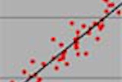
Moreover, the calcium scoring results were nearly identical (r=0.99, p < 0.0001). Algorithmic analysis showed "a very good correlation, we do not have any significant differences between the 80 kVp protocol and the 120 kVp protocol," Wintersperger said.
"To conclude, the combination of EKG pulsing and the 80 kVp protocol allows for adequate reduction of radiation exposure by 65%," he said. There is no significant impact upon the detection and quantification of the coronary artery calcifications, and basically the lower tube voltage should be recommended to reduce the radiation exposure from calcium quantification."
"How do you know it was the lower kVp and not the (EKG) pulsing that decreased your radiation?" came the question from the audience.
The group had tested this issue and found that about half of the dose savings came from tube modulation, and the other half from the lower kVp, Wintersperger said.
By Eric BarnesAuntMinnie.com staff writer
March 10, 2003
Related Reading
U.S. to evaluate whether x-rays should be labeled carcinogens, February 18, 2003
Waist size predicts optimal CT dose in virtual colonoscopy, February 7, 2003
Studies cast doubt on low-level radiation dangers, January 30, 2003
Worrisome radiation dose seen in CT lung screening, follow-up, December 23, 2002
Copyright © 2003 AuntMinnie.com