Echocardiographic determination of aortic flow propagation velocity (APV) can allow for bedside risk stratification of coronary artery disease (CAD), according to research published in the journal Medical Science Monitor.
"This novel parameter may be particularly useful in identifying individual patients who will benefit from further diagnostic strategies for CAD," wrote a research team from Yüzüncü Yıl University and Van Military Hospital, both in Van, Turkey.
The Turkish research team studied 91 patients with newly significant CAD found on coronary angiography and 36 patients with normal coronary arteries examined between June and October 2007. Exclusion criteria included acute myocardial infarction, creatinine level greater than 2 mg/dL or the need for dialysis, severe hepatic failure, aneurysm of the aorta, severe valvular heart disease, left ventricular ejection fraction (LVEF) less than 40%, atrial fibrillation, frequent premature beats, left bundle branch block, and inadequate echocardiographic image quality (Med Sci Monit, September 2008, Vol. 14:9, pp. MT42-46).
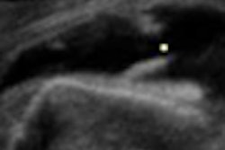
The echocardiography exam was performed at rest using a Vivid 3 echocardiography scanner (GE Healthcare, Chalfont St. Giles, U.K.) with a 3-MHz transducer by one of two experienced echocardiographers blinded to the clinical data and ongoing therapy. Aortic strain, aortic distensibility, aortofemoral pulse-wave propagation velocity (PWPV), and color M-mode propagation velocity of the descending aorta were then measured.
In the patient population, there were 23 cases of single-vessel CAD, 32 cases of two-vessel CAD, and 36 cases of three-vessel CAD. Seventy-three (80%) of the 91 patients with CAD were male and 36 (39.5%) were smokers, compared with 18 males (50%) and seven smokers (19.4%) in the patient group with normal coronary arteries. In other findings, the mean values of LVEF and APV were significantly lower with higher PWPV in patients with CAD compared with the control group (p = 0.001 and p < 0.001, respectively), according to the study team.
The researchers then performed multivariate regression analysis of the echocardiography variables, as well as age, hypertension, LDL cholesterol, diabetes, smoking habit, body mass index, and left ventricular ejection fraction. From this analysis, the researchers found that APV (beta = 0.850, p < 0.001) and PWPV (beta = 0.166, p = 0.008) were the only significant predictors of CAD.
"However, when APV was extracted from the regression model, the adjusted R2 decreased from 0.652 to 0.099," the authors wrote. "Therefore, APV is the main variable in the prediction of CAD. An APV model of ≤ 41 cm/sec, determined by receiver operating curve analysis, predicted CAD with 84.2% sensitivity and 97.2% specificity (positive predictive value: 98.7% and negative predictive value: 68.2%)."
The researchers also noted a significant correlation between APV and the severity of CAD (single vessel disease: r = -0.203, p = 0.022; two-vessel disease: r = -0.250, p = 0.005; three-vessel disease: r = -0.426, p < 0.001).
It might be expected that the decrease in the propagation velocity would not necessarily show coronary atherosclerosis due to the uneven distribution of atherosclerosis in different vascular territories, according to the researchers.
"However, we showed that APV predicted coronary atherosclerosis more powerfully than other methods of ultrasonographic aortic stiffness measurements," the authors wrote. "Furthermore, APV was the most significant and powerful predictor of CAD among the clinical and echocardiographic variables."
The researchers acknowledged several limitations of their study, including the reliability and reproducibility of the acquisition and reading of the different methods, the small size of the study population, and the relatively small number of control patients.
Nonetheless, the researchers concluded that measuring APV is a practical method for CAD risk assessment.
"Further studies searching for the effects of different cardiovascular risk factors, drugs, and treatment modalities on APV are needed," the authors wrote.
By Erik L. Ridley
AuntMinnie.com staff writers
September 16, 2008
Related Reading
Real-time 3D TEE helps guide heart-repair procedures, September 4, 2008
Quantitative stress echo can reliably find coronary artery disease, August 18, 2008
U.S. echo contrast agent use dips in Q2, August 14, 2008
FDA formally updates echo contrast black box warning, July 18, 2008
FDA ultrasound contrast safety meeting sparks discussion, hope for the future, June 10, 2008
Copyright © 2008 AuntMinnie.com