Improved imaging and therapeutic options have resulted in more congenital heart problems being diagnosed than in the past. Researchers in Germany believe they have found an easy, reliable way to deliver functional information in adult patients with complex congenital heart disease -- MRI.
Specifically, time-resolved contrast-enhanced MR angiography (MRA) of the thorax readily diagnoses adults with congenital heart disease quickly and reliably, according to Dr. Oliver Mohrs from Alice Hospital in Darmstadt, Germany; Dr. Steffen Petersen from the University of Oxford in the U.K., Dr. Thomas Voigtlaender from Cardiovascular Center Bethanien in Frankfurt, Germany; and colleagues.
For many patients who underwent surgical correction during childhood for congenital heart defects and have since reached adulthood, "the complex anatomy is not fully known, surgical reports are unavailable, and patients are lost to follow-up," the authors wrote.
"During adolescence, nonspecific clinical symptoms such as dyspnea, fatigue, or arrhythmias may develop. In contrast to neonates and children, in whom a wealth of information can be rapidly gained by echocardiography, older patients need a different diagnostic technique to reveal the underlying and surgical anatomy" (American Journal of Roentgenology, October 2006, Vol. 187, pp. 1107-1114).
MRI's utility is well-established for both diagnostic and functional imaging, though keeping imaging time to a minimum requires knowing the underlying diagnosis before specific functional tests are conducted, the authors wrote. Time-resolved contrast-enhanced MRA, which combines tomographic and projection imaging methods, is a fast way to obtain the maximum information very quickly during a single procedure.
The study sought to evaluate the diagnostic accuracy of time-resolved contrast-enhanced MRA of the thorax in adult patients with known or suspected complex congenital heart disease.
The researchers examined 20 patients (mean age, 38 ± 14 years; range, 16-73 years) with congenital heart disease who underwent contrast-enhanced turbo fast low-angle shot MRA. The radiologists acquired 30 consecutive coronal 3D slabs with a one-second frame rate; the mask (first dataset) was subtracted from subsequent images.
Image quality was evaluated on a five-point scale (1, not assessable; 5, excellent image quality), and the images were examined for 12 diagnostic parameters. Each parameter yielded one point for correct diagnosis, summarized into three categories including anatomy of the main thoracic vessels, sequential cardiac anatomy, and shunt detection.
The results were compared with a combined clinical reference comprising medical or surgical reports and other imaging studies, all of which served as the reference standard.
The results showed a mean image quality of 3.7 ± 1.0, the authors wrote, and 220 of 240 (92%) of the single diagnostic parameters could be analyzed using a binary approach.
 |
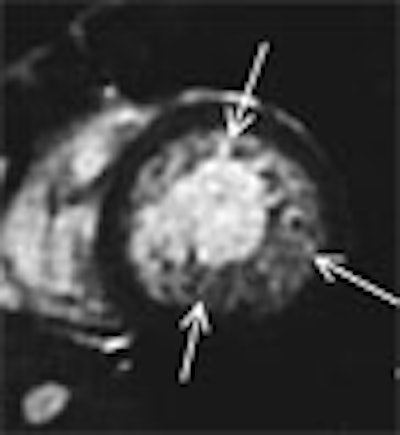
| Fig. A (above), 23-year-old woman referred to MRI with suspicion of transposition of great arteries because of prominent trabeculation of subaortic ventricle in echocardiography. Time-resolved coronal maximum-intensity-projection angiograms (B-C, below) and cine short-axis view (D, bottom). The angiograms show normal time course of enhancement of subpulmonary atrium and ventricle and pulmonary arteries (A); and enhancement of pulmonary veins, subaortic atrium, ventricle, and aorta (B). Note decreasing enhancement of subpulmonary ventricle from A to C, indicating absence of relevant left-to-right shunt flow. This patient suffered from noncompaction myocardium, which is visualized on time-resolved MR angiography (C) (arrow) and is shown on cine short-axis view (D) (arrows). Republished with permission of the American Roentgen Ray Society from AJR 2006; 187:1107-1114. |
 |
 |
 |
"The percentage of maximum diagnostic points, the sensitivity, the specificity, and the positive and the negative predictive values were all 100% for the anatomy of the main thoracic vessels; 97%, 87%, 100%, 100%, and 96% for sequential cardiac anatomy; and 93%, 93%, 92%, 88%, and 96% for shunt detection," Mohrs and his study team wrote.
Several MRI techniques can be used to visualize and quantify congenital cardiac abnormalities, but the drawback of most techniques is their need to focus on a specific finding due to time constraints, they explained.
In contrast, time-resolved MR offers a fast overview at the beginning of the MRI exam, which enables "further specific morphologic and functional analysis during the same session," they wrote.
The technique yielded good image quality (3.7/5.0) and reliable diagnosis. Diagnostic accuracy was only slightly reduced in detecting shunt flow. Pulmonary regurgitation presented a pitfall, "implicating a plateau phenomenon in the subpulmonary ventricle," the authors wrote
Some structures were not evaluable because of partial amputation at the edge of the slab, they cautioned. There was a trade-off between sufficient temporal resolution of the 3D datasets with the number of slices acquired. Further developments in parallel imaging techniques might help to hasten the data acquisition and improve the abililty to assess peripheral structures, they wrote.
"In our opinion, based on initial experience, the amount of contrast agent should be as small as possible using a high flow rate to obtain a compact bolus," the group wrote. "The compactness of the bolus is mandatory to separate the filling times of the different vascular or cardiac compartments. We used a mean dosage of 0.07 ± 0.02 mmol/kg/body weight of gadopentetate dimeglumine to achieve such a compact bolus of contrast agent without a major loss of vessel visualization. In addition, a second injection for high-resolution MR angiography with a sufficient signal-to-noise ratio is possible if needed during the same examination."
Time-resolved contrast-enhanced MRA offers "rapid anatomic and functional information in adult patients with complex congenital heart disease in a single breath-hold," the researchers concluded. Its high accuracy may allow physicians to tailor specific MRI sequences later in the same session.
By Eric Barnes
AuntMinnie.com staff writer
October 18, 2006
Related Reading
MR angiography offers greater detail in vascular imaging, March 10, 2006
Studies showcase MRA at its best in arteries and combined with CAD, November 17, 2005
MRA shows good results in detecting coronary stenosis, March 7, 2005
3D contrast MRA may be optimal for evaluating aortic dissection, December 30, 2004
Whole-body MR permits rapid evaluation of lower peripheral arterial system, July 16, 2004
Copyright © 2006 AuntMinnie.com