SEATTLE - SPECT may be the clinical standard for perfusion assessment of myocardial ischemia and PET may offer more accuracy, but MRI is an equally valid option, according to a presentation Sunday at the International Society for Magnetic Resonance in Medicine (ISMRM) meeting.
"Myocardial ischemia occurs when there is insufficient blood flow, or perfusion, to the myocardium. This results in inadequate delivery of oxygen," said Frederick Epstein, Ph.D., an associate professor of radiology and biomedical engineering at the University of Virginia in Charlottesville. "Numerous modalities can be used for imaging perfusion defects…. MRI has at least three different methods that can be used for myocardial perfusion."
Epstein outlined the specifics of the most popular technique, first-pass MRI, and also touched on more experimental MR methods. He also discussed the off-label use of contrast agents and disclosed that Epix Pharmaceuticals of Cambridge, MA, as well as Siemens Medical Solutions of Malvern, PA, support his research.
First-pass MRI
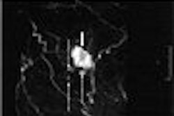
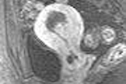
This method is based on tracer kinetics, using rapid T1-weighted imaging to detect the first passage of a gadolinium bolus as it flows through the left ventricular cavity and myocardial microcirculation. The contrast agent is T1-shortening, resulting in a relatively high spatial resolution (low spatial resolution remains a pitfall of SPECT). First-pass MRI can also distinguish transmural differences in perfusion.
First-pass MRI is typically done during stress and at rest. Stress-induced demand ischemia is a potential cause of a lack of contrast enhancement, but Epstein cautioned that there might be other reasons for no enhancement, such as myocardial infarction or artifact.
"The differential can usually be clarified by the acquisition of additional stress first-pass images and by acquiring inversion-recovery images to detected delayed hyperenhancement," he said.
For true demand-ischemia, lack of contrast enhancement will be seen at stress on first-pass images but not at rest, Epstein added. Delayed hyperenhancement is not seen on inversion recovery images.
First-pass MR pulse sequences
Saturation-recovery fast gradient echo (TurboFLASH) is generally considered the optimal pulse sequence, Epstein said, but imaging experts should consider other sequences as well. Hybrid fast gradient-recalled echo and echo-planar imaging (GRE-EPI) can acquire data more rapidly, resulting in greater slice coverage and less cardiac motion. Steady-state free precession (SSFP) generates greater contrast, which means higher sensitivity for detecting perfusion defects.
With TurboFLASH, "image acquisition time is about 200 milliseconds, or two to three slices per heartbeat can be covered, particularly at stress," Epstein said. With GRE-EPI, "instead of single echo, a short train of echo is required … roughly (doubling) acquisition to four to five slices per heartbeat." Finally, SSFP will increase the signal-to-noise ratio (SNR) but will not increase acquisition speed, he added.
Epstein pointed out that GRE-EPI and SSFP are still proving themselves. Recent research has yielded conflicting results, he said. In a 2005 paper in Radiology that compared the two sequences, TurboFLASH came out the winner; in another 2005 paper that compared GRE-EPI to SSFP, the latter was favored (Radiology, April 2005, Vol. 235:1, pp. 237-243; Magnetic Resonance in Medicine, November 2005, Vol. 54:5, pp. 1123-1129).
Session moderator Dr. Scott Flamm asked Epstein if he would hazard a guess as to which sequence would come out the winner. Epstein reiterated that it was still too soon to tell, but that better temporal resolution and fast acquisition time with less heart movement were certainly desirable.
Quantitative analysis
Images can be analyzed qualitatively or visually, but there are also components of doing quantitative analysis, according to Epstein. "What you do is put down a region of interest (ROI) … and generate the time-intensity curve," he said.
Once time-intensity curves are generated, quantitative model-based deconvolution methods are applied to estimate perfusion in units of mL/g/min or by solving modified Kety equations. For both of these methods, estimates of input function (signal intensity in the myocardium) and tissue function (signal intensity in the left ventricular blood pool) are required (Medical Physics, January 1998, Vol. 25:1, pp. 73-84; Magnetic Resonance in Medicine, September 1993, Vol. 30:3, pp. 337-346).
"The question here is to quantify or not to quantify?" Epstein asked. He explained that quantification is a time-consuming process, especially if the patient's breath-hold technique is poor, which requires adjustments to the contours of the heart. But take the time to quantify results in improved agreement with microsphere measurements and improved accuracy, he said.
Methods that utilize the input function for image analysis generally require contrast agent, but in what concentration? A contrast dose of 0.05-0.15 mmol/kg could produce good enhancement -- and more false positives. Epstein recommended a lower dose (0.025 mmol/kg) because the signal from the left ventricular cavity is not linear with contrast agent concentration. This will limit the SNR of the myocardium during the first pass of contrast medium, Epstein warned.
More sophisticated contrast administration, such as dual bolus and dual contrast sequences, are in the works. In the latter, a relatively high contrast concentration is used for multislice imaging, he said.
"The addition of delayed contrast-enhanced MR distinguishes myocardial infarction ischemia," he said.
Clinical studies, future directions
Early studies put the sensitivity of first-pass MRI in ischemia at 65% to 92%. Specificity ranged from 75% to 100%. A more recent study indicated a higher area under the receiver-operator curve for first-pass MRI in comparison to SPECT (Circulation, May 8, 2001, Vol. 103:18, pp. 2240-2235; Radiology, October 2003, Vol. 229:1, pp. 209-216).
In the more immediate future, blood oxygen level-dependent (BOLD) imaging and arterial spin labeling (ASL) offer other MRI methods. Using BOLD means looking for different magnetic properties in either oxygenated or deoxygenated blood, resulting in altered signal intensity. ASL measures the T1-weighted recovery of myocardium.
Finally, the advantage of cardiac MR at 3 tesla is that it provides higher SNR to 1.5 tesla, but it also may generate more artifacts, Epstein said.
Session moderator Dr. Joao Lima told the audience: "If I were to advise anyone beginning their career in MR, I would advise that they 'go perfusion.' That's where the money is."
By Shalmali Pal
AuntMinnie.com staff writer
May 8, 2006
Related Reading
First-pass MR detects subtle ischemia, February 21, 2006
MRI differentiates AMI from myocarditis, November 21, 2006
Study quantifies prevalence of noncardiac findings on cardiac MR, January 25, 2005
Stress MRI tops echo for coronary disease detection, May 26, 2004
Myocardial perfusion measured by MRI useful in detecting heart disease, August 4, 2003
Copyright © 2006 AuntMinnie.com