Attenuation values in coronary calcium scoring can vary significantly depending on the scanner, and even by patient body type, according to a new study in Radiology based on nearly 10,000 participants in two major trials. Fortunately, the researchers proposed a potential solution in the form of calibration phantoms that could improve accuracy in diagnosis and follow-up.
"Many scanner and participant factors (e.g., the anatomy of interest, x-ray beam energy, beam hardening) interact in complex ways to determine the attenuation," wrote Jennifer Clark Nelson, Ph.D.; Richard A. Kronmal, Ph.D.; Dr. J. Jeffrey Carr; and colleagues. "Thus, image attenuation may differ between CT scanners, scanning protocols, and participants over time. In turn, CAC (coronary artery calcium) measurements can be highly affected by image attenuation because they depend on the detection of aggregates of contiguous image pixels with attenuation values greater than 130 HU and because some CAC measures (e.g., the Agatston score) depend on present attenuation thresholds.... Hence, variability in image attenuation increases measurement error and reduces the accuracy and precision of CAC quantification" (Radiology, May 2005, Vol. 235:3, pp. 403-414).
The researchers are from several institutions including the University of Washington in Seattle; Wake-Forest University School of Medicine in Winston-Salem, NC; and the University of California at Los Angeles and Irvine.
For their study, the researchers performed a standardized scanning protocol at all of the sites that relied on data from two earlier heart scanning trials: the Coronary Artery Risk Development in Young Adults (CARDIA) trial, and the Multi-Ethnic Study of Atherosclerosis (MESA). The Radiology authors were also given access to the patient data from the two earlier trials for use in their phantom study.
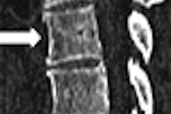
Two anthropomorphic phantoms, a calibration and a torso phantom, were scanned at each site. The calibration phantom contained four rods of known calcium hydroxyapatite concentrations (0, 50, 100, and 200 mg/mL). The phantom was positioned beneath each participant so that its length covered the expected length of the heart, and it was scanned in every section. The torso phantom contained a single rod with 100 mg/mL of calcium hydroxyapatite, and was scanned for quality-control purposes every two to four weeks, the authors wrote.
Leveling the playing field
The group developed an attenuation-adjustment procedure to reduce variability across scanners and participants, and a method for calibrating image attenuation to a standard level so that CAC measurements could be made on images with comparable attenuation levels.
This process involved using a representative image sample to define standard attenuation levels, and calculating it to the other images. Then the average attenuations corresponding to known calcium quantities were used as standards. After measuring the attenuation in the four calibration rods of the phantoms, a formula was devised to correct the attenuation between the observed CT values and the standardized values. Calibrations were also calculated for each participant. Finally, the behavior of calibration phantom data was used to develop a calibration algorithm.
"We calibrated CT scans acquired in 3,041 participants in the CARDIA study at the year-15 examination ... and CT scans acquired in 6,814 participants in the MESA at the baseline examination," the authors wrote. "The calibration algorithm is simple and was easily programmed into the computer reading software at the CT reading center, so that attenuation adjustments were automatic.... Most importantly ... the calibration method yielded the desired effect of more comparable CAC measurements between images obtained with different scanners and in different participants."
The results showed that mean CT attenuation values of a standard calibration phantom scanned beneath participants varied significantly according to scanner (p < 0.01), as well as for the participant's body mass index (BMI) (p < 0.01). The lowest values were obtained for the Siemens' multidetector-row CT scanners (110 HU) (Siemens Medical Solutions, Malvern, PA) followed by GE's Imatron electron-beam scanners (116 HU) (GE Healthcare, Chalfont St. Giles, U.K.) and GE's LightSpeed multidetector-row scanners CT (121.5 HU).
"These differences are not surprising because the CT study protocol involved the use of different peak voltages with different scanners, and higher-energy beams tend to undergo lesser degrees of attenuation," the authors wrote. "However, beam energy does not account for all the variability between scanners, as evidenced by the 9.1 HU difference between electron-beam CT systems (all of which scanned at 130 kVp). This is likely driven by scanner calibration differences."
The participant's body size also affected image attenuation significantly, with the lowest attenuation levels recorded among obese participants and the highest levels seen in normal-weight or underweight participants.
"Values were ... lower for morbidly obese (BMI ≥ 40.0 kg/m2) participants (108.9 HU), followed by obese (BMI 30.0-39.9 kg/m2) (114.8 HU), overweight (BMI 25.0-29.9 kg/m2) (118.5 HU), and normal-weight or underweight (BMI < 25.0 kg/m2) (120.1 HU) participants," the authors stated. "Agatston score calibration adjustments ranged from -650 to 1,071 (mean -8 ± 50 [standard deviation])."
The inverse association between BMI and attenuation has been seen by other researchers, and may be due to hardening of the x-ray beam as it passes through tissues proximal to the anatomy of interest, the researchers noted.
Interpreting the results
"These scanner and participant attenuation differences are a matter of concern because methods for detecting and measuring CAC inherently depend on image pixels exceeding preset attenuation thresholds," they wrote. "Thus, attenuation variability can cause differences in CAC measurements that are not due to true disparities in CAC burden between participants."
The postcalibration attenuation adjustments were small on average, they noted, although some individual Agatston score adjustments were large (up to 1071), particularly among individuals with large unadjusted Agatston scores. Some adjustments resulting from automated calibration changed the clinical categorization of coronary calcium burden, they added.
The data also suggest that attenuation adjustments due to body size should be performed for all examinations. Still, they cautioned, "the physics of x-ray imaging are complex, and our use of a phantom that is placed relatively distant from the coronary arteries may not result in appropriate measurement of attenuation in the coronary tree in the same image section."
More research is needed before a strong case could be made for adjustments on body size alone. "We have shown, however, that image attenuation, and therefore the meaning of the Agatston score, is not the same from scanner to scanner and from participant to participant," they wrote. "Research studies that involve the use of different scanner technologies would most likely benefit from the utilization of attenuation adjustment."
The differences were especially large when different kilovoltage settings were applied.
More data are also needed to evaluate the potential benefit of attenuation-adjusted CAC measurements, including morbidity and mortality follow-up data, the group wrote.
By Eric Barnes
AuntMinnie.com staff writer
May 31, 2005
Related Reading
Coronary calcium predicts brain lesions, cognitive decline in the elderly, April 28, 2005
As cardiac CT interest heats up, so does EBCT, MDCT debate, March 31, 2005
Aortic valve calcification equals aortic stenosis, December 1, 2004
Most MDCT scanners fall short in cardiac imaging, September 3, 2004
Coronary artery calcium detected by tomography may predict heart disease, May 15, 2003
Copyright © 2005 AuntMinnie.com