Total knee arthroplasty has been around for decades, but only recently have minimally invasive surgical techniques, space-age knee replacement materials, and advanced imaging techniques improved the results dramatically.
Still it's a tricky undertaking. Accurate implant alignment and ligament balance are necessary, and because conventional radiography can be inadequate, CT is taking on a bigger role in presurgical planning, with increasing use of 3D measurements. But a group of Korean researchers wondered if measurements based on 3D reconstructions were accurate and reproducible.
"For successful total knee arthroplasty, accurate implant alignment and ligament balance are essential," wrote Drs. In Sook Lee, Jung-Ah Choi, and colleagues from Seoul National University and the affiliated Bundang Hospital in Seoul, South Korea. "The dimension of the distal femur and proper rotational alignment of the femoral component are especially critical for the outcome of total knee arthroplasty. The importance of correct sizing of components for total knee arthroplasty for optimal function and long-term results has been stressed in many reports."
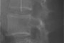
Quantifying the shape of bones in the osteoarthritic knee is particularly difficult due to knee deformity, and the size can be measured differently depending on patient position, the group wrote in the June issue of American Journal of Roentgenology (Vol. 186:6, pp. 1,778-1,782).
The study sought to determine the consistency of measurements of the distal femoral condyle on 16-slice multidetector-row CT (MDCT). Over a two-year period, 33 patients (two men, 31 women; age 53-89, mean age 71) with osteoarthritis underwent MDCT scans (Mx8000 IDT, Philips Medical Systems, Andover, MA) for preoperative planning of total knee arthroplasty.
Images were acquired at a 2-mm slice thickness and 1-mm interslice gap, with matrix size of 512 x 512. Images were loaded onto a Rapidia 2.8 workstation (Infinitt, Seoul, South Korea), and only portions of the distal femoral condyle were reformatted with 3D reconstructions, according to the authors.
Prospective analysis on the workstation included measurements of transepicondylar distance, maximum anteroposterior dimension of medial and lateral femoral condyles, and trochlear width, measured using multiplanar reconstructions and 3D volume-rendered images.
"For accuracy, dot cursors for measuring on the 3D volume-rendered image were placed in identical positions on corresponding axial, coronal, and sagittal images," they wrote.
Two radiologists independently read the results and compared the degree of agreement, but because their results were nearly identical, only those of one radiologist were compared with the interoperative findings, which were measured with a caliper.
Then, to test the reliability of the measurements, the group repeated them on a CT workstation after two months and compared them to previous values.
"The mean values of transepicondylar distance, maximum anteroposterior dimension of medial and lateral femoral condyles, and trochlear width were 75, 57, 58, and 38 mm at first measurement; 76, 58, 59, and 39 at second measurement on the CT workstation; and 79, 57, 60 and 42 mm at intraoperative measurement, respectively," the group wrote.
In the reliability analysis between the first and second measurements on the workstation and the intraoperative measurements, the kappa values were 0.84 for the transepicondylar distance, 0.81 for the maximum anteroposterior dimension of the medial femoral condyle, 0.89 for the maximum anteroposterior dimension of the lateral femoral condyle, and 0.62 for the trochlear width. For the second measurement kappa values were 0.86, 0.77, 0.84, and 0.61, respectively.
"Intraoperative measurements and measured values on the CT workstation showed excellent and almost perfect agreement, and intraobserver agreement was almost perfect," they wrote.
Still, in patients with severely deformed knees and large or multiple osteophytes, the differences between MDCT and interoperative measurements tended to be larger, and therefore "more careful measurements and averaging of repeated measured values would be needed in such cases," the group noted. "However, some discrepancy may simply be due to the inclusion of soft tissue in the interoperative measurements, rather than from the inaccuracy inherent to measurement on CT."
"Previous studies showed a difference in sizing of the distal femur for total knee arthroplasty according to the patient's position and rotational alignment on conventional radiography, MRI, and conventional CT," Lee and colleagues wrote.
With the aid of MDCT and an advanced workstation, "we could measure the proper size of the femoral component on 3D reconstruction, multiplanar reconstruction coronal and sagittal images, and axial images, irrespective of rotational alignment," they wrote.
"Reliable preoperative 3D measurements with 3D reconstructions using 16-MDCT can be applicable in several instances, especially in the field of computer-assisted surgery, for which its use needs further investigation," the group noted.
By Eric Barnes
AuntMinnie.com staff writer
May 31, 2006
Related Reading
Hospital for Special Surgery's Pavlov discusses orthopedic radiology's future, February 17, 2006
X-ray protocol improves total knee arthroplasty follow-up, March 26, 2004
Copyright © 2006 AuntMinnie.com