AUSTIN, TX - The rapid evolution of modalities used in the practice of radiology, in particular multislice CT scanners, is fast producing a daily volume of images that may jeopardize the capacity of radiologists to interpret them, according to a presentation at the Society for Computer Applications in Radiology (SCAR) annual meeting.
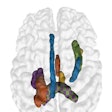
As the number of images to be interpreted on a daily basis has increased, the radiology community has begun to evolve strategies for handling the workflow, among them the use of 3D imaging, said Dr. Eliot Siegel.
Siegel, professor and vice chairman of information systems in the department of diagnostic radiology at the University of Maryland School of Medicine in Baltimore, and chief of imaging at the Veterans Affairs Maryland Health Care System, shared his thoughts on maximizing advanced image processing at the meeting on Friday.
According to Siegel, there are currently three models for advanced image processing, such as 3D imaging and multiplanar reconstruction (MPR):
- Reconstruction can be performed at the CT scanner.
- Reconstruction can be done dynamically by the radiologist.
- Reconstruction can be accomplished in a dedicated 3D lab.
The most popular model, he said, is to have a technologist conduct the advanced image processing at the scanner. This is highly inefficient, as it greatly reduces the amount of patient throughput on the system. In addition, Siegel noted, the technologist performing the reconstruction may or may not know the preferences of the radiologist or referring clinician.
Providing the capability for radiologists to dynamically perform advanced image processing on their workstations mitigates the preferences limitation that can exist when CT technologists construct these image sets on scanner workstations. However, the reconstructions take valuable diagnostic interpretation time away from radiologists as they wait for the image datasets to render.
"The best model given the current reimbursement environment and combination of routine and complex types of image reconstructions and visualizations is probably a combination of 3D lab (real or virtual) and advanced radiologist/clinician workstation," Siegel said.
The advantages to this workflow structure are that it allows technologists to scan and then send the images to an advanced image processing lab for processing, without interrupting patient throughput in a scanner or compromising a radiologist's diagnostic image interpretation time, he said. The workflow structure also enables the creation of a subspecialist with expertise in advanced image processing, which can increase throughput in this area. It can generate additional income for the practice as well.
Although reimbursement varies for 3D imaging studies according to the type of study and the region of the U.S. in which it is performed, having this capability may add studies to a practice, according to Siegel. One reason is that once referring clinicians are exposed to these studies, they tend to order them in greater volume, he noted.
Setting up a 3D lab requires more than setting up dedicated workstations, Siegel said. Dr. Peter Janick of Lansing Radiology Associates in Lansing, MI, has offered leadership advice for practices interested in developing an advanced image processing lab, which Siegel cited during his presentation.
Janick believes that a strong radiologist and technologist experienced at operating the workstation are needed to lead the process. This provides an expert voice at the beginning of the development process, as well as establishing an accessible individual for technologists and clinicians to contact during the inception of the service.
It helps to have a "pioneering spirit" to try new applications and tough cases, according to Janick. It is also prudent to set aside dedicated time to both read and innovate cases and protocols to master the technology.
But there are pitfalls that radiologists should be aware of as they venture into the advanced processing arena, Siegel cautioned. No standards exist across vendors for presentation, and Siegel said that he has found variability in quantitative measurements from workstation to workstation. He counseled that radiologists may have to conduct a "reality check" on numbers generated by these workstations.
Siegel also noted that, in his experience, bone removal software may remove portions of the vessel resulting in inaccurate assessment of stenosis or other pathology. Finally, he suggested that advanced image processing adopters need to be careful to balance scientific accuracy with the artistic creativity possible with the technology.
By Jonathan S. Batchelor
AuntMinnie.com staff writer
April 30, 2006
Related Reading
Technologists take advantage of 3D opportunity, April 25, 2006
Expanded role looms for 3D in breast imaging, April 11, 2006
Setting up a cutting edge 3D lab, April 7, 2006
Technological advances drive new 3D CPT codes, February 21, 2006
How to find and train a 3D technologist, June 17, 2005
Copyright © 2006 AuntMinnie.com
