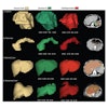
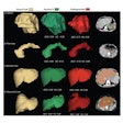
ORLANDO, FL - An important component of tracking disease progression is the comparison of patient images from older studies with their latest images, especially in CT, MR, and PET exams. Image matching is time-consuming work, however, and poor alignment can lead to uncertainty and error on the part of the interpreting radiologist, according to a presentation this morning at the Society for Computer Applications in Radiology (SCAR).
Dr. Bradley Erickson, chair of SCAR's annual meeting program committee and an associate professor in radiology and medical informatics at the Mayo Clinic in Rochester, MN, shared the results of a study on the impact of automated image alignment on image interpretation conducted at his institution.
"We prospectively recruited 20 sequential CT or MR examinations that had comparison examinations for both neurological and non-neurological body parts -- 20 in each of the two categories -- for our automated image alignment study," Erickson said.
The researchers set out to determine whether the use of automated image alignment improved report generation time and image interpretation. For purposes of the study, one index pathology was selected for reporting purposes, he said.
The group used the Insight Segmentation and Registration Toolkit (ITK) open-source application, sponsored by the National Library of Medicine, to perform the automated image alignment and displayed the results on a Siemens Medical Solutions (Malvern, PA) lab PACS. Both registered (autoaligned) and nonregistered mages were interpreted by a team of six radiologists (three neuroradiologists and three body radiologists) at the Mayo Clinic.
"Our original intent was to conduct a multisite study, but this didn't happen," Erickson said. "Due to high workloads in radiology, we had difficulty in recruiting readers from other institutions. In addition, some prospective sites were resistant to research being loaded onto a clinical PACS."
Automated image alignment took a mean of 68 seconds of computer time, and 98% of the studies were able to register alignment, Erickson said.
Erickson found that the time to report a registered neurological study was 82 seconds, compared with a nonregistered neurological exam reporting time of 113 seconds. The team reported that the time to report a registered body study was 85 seconds, compared to the nonregistered body study reporting time of 95 seconds.
Overall, Erickson said, the use of automated image alignment yielded report generation time savings of approximately 25%. In terms of report agreement, he said that there was an overall kappa of 0.52 for the registered image reports, compared with 0.46 for the unregistered image reports.
"The radiologists who used the automated image alignment application all felt that the tool would make their practice better and more efficient," he said.
Erickson noted that the group discovered that 78% of the neurological MR exams and 92% of the neurological and body CT exams were an easy match with the ITK software. Body MR exams, however, proved slightly more difficult to register, with only 52% of these studies reported as an easy match.
They also found that standard scanning protocols are very important to the success of automated image alignment, as series with a high similarity (those with only one series of a different protocol) were more easily interpreted and reported than those of low similarity. In addition, Erickson said, the application appears to be better suited for rigid structures.
"In body CT, the change in breath and motion diminished the effectiveness of automated image alignment," he said. "However, with the advent of higher-speed CT technology, this may be eliminated."
By Jonathan S. Batchelor
AuntMinnie.com staff writer
June 2, 2005
Related Reading
Tibial-talar ratio on x-ray reliably shows ankle alignment, May 6, 2005
More PET/CT lung masses seen with hardware-based fusion, April 26, 2005
Lung nodule analysis makes progress, not perfection, March 5, 2004
Auto co-registration of MR, SPECT images could avoid prostatectomy, June 6, 2000
Fused Image Tomography: An Integrating Force, August 1, 1999
Copyright © 2005 AuntMinnie.com
