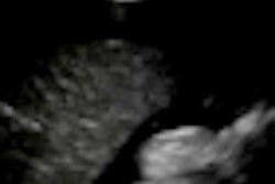
Thanks to new perfusion imaging techniques, CEUS guidance for targeted biopsy could substantially increase the detection rate for positive biopsy cores compared with systematic biopsy approaches, said lead author Peter Frinking, PhD, of Bracco Imaging.
The team sought to develop a new approach for improved prostate cancer detection in prostate peripheral zone tissue, and compare the technique against histopathology of whole-mount prostatectomy samples, Frinking said.
The group's perfusion imaging approach is based on a statistical analysis of parametric images of wash-in rate, which is defined as the ratio of peak enhancement and rise time, according to the authors. This perfusion parameter reflects the rate of contrast enhancement.
They tested the method on contrast-enhanced sequences from 20 patients who were scanned on an iU22 (Philips Healthcare) scanner after 2.4-mL bolus injections of SonoVue (Bracco).
Color-coded probability maps generated from the statistical analysis of wash-in rates led to considerable improvements in sensitivity and specificity over standard time-intensity curve (TIC) analyses in the same areas, according to the researchers.


