Dr. Christopher Maroules from UT Southwestern Medical Center in Dallas is scheduled to present results from a study that evaluated 10 healthy volunteers and 10 patients with pulmonary arterial hypertension.
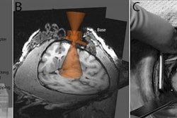
The MRI protocol included 2D flow alternating inversion recovery with an extra radiofrequency pulse (2D ASL-FAIRER) along with 2D phase-contrast imaging of the pulmonary arteries to measure pulmonary artery flow. In addition, 10 subjects underwent a second 2D ASL-FAIRER scan to help determine reproducibility.
Two-dimensional ASL-FAIRER achieved high interobserver reproducibility and moderate test-retest reproducibility for regional lung perfusion. Maroules and colleagues also found good correlation between perfusion in the right lower lobe and pulmonary artery flow, but weaker correlation between perfusion in the right upper lobe and pulmonary artery flow.
In addition, the relative dispersion of lung perfusion was greater among patients with pulmonary arterial hypertension (1.25 ± 0.41), compared with the healthy volunteers (0.93 ± 0.32, p = 0.06), but the difference was deemed not statistically significant.
Noncontrast 2D ASL-FAIRER measurement of regional lung perfusion at 3 tesla is "feasible and demonstrates good reproducibility," the group concluded. "ASL-derived regional lung perfusion correlates with phase contrast pulmonary artery flow."