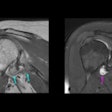
That conclusion comes from researchers at the Medical University of Vienna, who say the results show that DTI will help optimize surgical procedures, because it will detail preoperative 3D visualization and characterization of peripheral nerve involvement in benign and malignant soft-tissue tumors.
"The advantage of MRI in general is the excellent tissue contrast and the clear delineation of peripheral nerves in vivo," said study co-author Gregor Kasprian, MD. "The excellent signal-to-noise ratio permits the application of diffusion-tensor imaging as a new interesting imaging technique. Postprocessing of the DTI original data allows the 3D visualization of peripheral nerves and provides the opportunity to acquire quantitative imaging data," he said.
The study reviewed 27 patients (16 females, 11 males; mean age, 54.9 years) with suspected soft-tissue tumors, including 13 cases of sarcoma, four patients with peripheral nerve sheet tumors, three cases of benign tumors, and two patients each with myxoma, lipoma, and fibroma.
All 27 patients were examined using a 3-tesla MRI system and an echo-planar DTI sequence in addition to the standard diagnostic protocol. Software was used to define multiple regions-of-interest (ROI) along major and potentially affected peripheral nerves, and tractography was performed. Fractional anisotropy values were measured within lesional and nonlesional ROI.
The researchers found that tractography successfully depicted the 3D course of the sciatic nerve in 12 patients, the tibial nerve in seven cases, the femoral nerve in four cases, the peroneal nerve in two patients, and the median and musculocutaneous nerves in two cases each.
In 18 cases showing an immediate anatomical relationship with the soft-tissue tumor, the nerves appeared continuous in nine cases or partially discontinuous in six patients. In two of the three cases with complete nerve discontinuity, nerve infiltration was histologically proven.
"The initial results in our study indicate a potential use to identify nerve infiltration of peripheral nerves in soft-tissue tumors," said Kasprian, who will present the study in Chicago. "Thus, the clinically important question -- whether peripheral nerves are involved by the malignant lesions -- can be answered. This helps in further planning of the surgical therapy of these cases and may allow a limb-salvaging approach, if nerve involvement can be excluded."
Kasprian and colleagues plan to continue their research in this area, using DTI to differentiate between neurofibromas and schwannomas. "Moreover, further surgical correlation should provide important data on the validity of our findings," Kasprian added.


