The effectiveness of medical imaging is directly dependent upon a radiologist's ability to search through an image set, recognize abnormalities, and contextualize those abnormalities with respect to other information regarding the health status and past medical history of the patient, according to presenter Dr. Geoffrey Rubin of the Duke Clinical Research Institute.
"At this time, much of this process is opaque as a part of the perceptual and cognitive toolkits that we all use to accomplish our activities of daily life," Rubin told AuntMinnie.com. "While radiologists undergo extensive training to interpret medical images, little is known regarding how they use their innate perceptual and cognitive characteristics to interpret images."
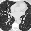
The research team used eye-tracking techniques and a log of mouse-driven scrolling activity to record 13 radiologists as they interpreted 40 lung CT scans that contained between three and five synthetic nodules.
"We find a remarkable variation in the extent to which radiologists are able to detect nodules with their peripheral vision and how frequently they miss nodules that are in the center of their gaze," Rubin said. "Our study provides a basis for characterizing a radiologist's gaze as a moving probability cloud that characterizes the momentary likelihood that a nodule will be detected or not. From this work, we may one day be able to enhance the reading environment to optimize detection based on the tendencies of individual radiologists as well as use the principles discovered to enhance radiologist training."
Learn more by sitting in on this Thursday morning presentation.


