By Eric Barnes, AuntMinnie.com staff writer
    November 6, 2012

    RSNA 2012 caps an extraordinary year for CT fans who like their technology high and their radiation doses low. The CT-focused scientific sessions at this year's show -- a few dozen of which are highlighted below -- explore a broad array of CT topics that are driven, more than anything, by the need to get more image information with less radiation.

    Scanning with different x-ray energies has really blossomed this year thanks to new, more adaptable CT hardware and software that brings the data to life. To note a couple of the many presentations on this topic, researchers from the University of Michigan are using high-kV virtual monochromatic images to get a better look at spine hardware (Wednesday, November 28, 11:10 a.m.-11:20 a.m., SSK11-05, Room N230).

    Meanwhile, a Harvard team is using multienergy CT to boost the accuracy of its virtual colonoscopy computer-aided detection (CAD) software (Tuesday, November 27, 3:50 p.m.-4:00 p.m., SSJ08-06, Room E353C). And Italian researchers are using iodine quantification to better characterize renal masses (Friday, November 30, 11:30 a.m.-11:40 a.m., SST07-07, Room E351).

    Road to RSNA 2012: CT Preview Dose reduction also shares top billing, of course, and each successive RSNA show highlights more ways to make it happen. For example, a team from China is using gated dual-source CT angiography to scan even atrial fibrillation patients at low doses (Monday, November 26, 10:30 a.m.-10:40 a.m., SSC01-01, Room S405AB). Next-generation iterative reconstruction is becoming standard practice, with vendors' offerings from model-based iterative reconstruction (Monday, November 26, 11:00 a.m.-11:10 a.m., SSC05-04, Room E353A) to AIDR 3D (Wednesday, November 28, 3:50 p.m.-4:00 p.m., SSM20-06, Room S404AB) getting a workout.

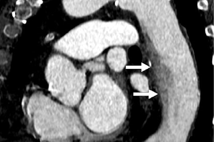
    New photon-counting CT detectors are making better use of energy information, offering big opportunities for dose reduction and, in one case, separating vulnerable from stable coronary artery plaque (Monday, November 26, 3:10 p.m.-3:20 p.m., SSE21-02, Room S403A). Faster scanners are cutting contrast doses too (Monday, November 26, 3:10 p.m.-3:20 p.m., SSE05-02, Room S404CD).

    In cardiac imaging, what's the reason behind higher rates of myocardial infarction among blacks? Researchers from the Medical University of South Carolina take a stab at the question (Monday, November 26, 11:40 a.m.-11: 50 a.m., SSC02-08, Room S504AB). And a Johns Hopkins University team is getting anatomic, functional, and motion information out of a single 64-detector-row coronary CT angiography (CTA) scan (Wednesday, November 28, 3:40 p.m.-3:50 p.m., SSM03-05, Room S502AB).

    And you won't want to miss the results of ROMICAT II, demonstrating that coronary CTA cuts hospital time and leaves patients better off than standard care (Wednesday, November 28, 8:30 a.m.-8:45 a.m., VSCA41-01, Room S502AB).

    But wait, there's more: Characterizing emphysema in patients with chronic obstructive pulmonary disorder has made considerable progress this year; one group is using low-attenuating clusters to identify affected regions (Monday, November 26, 10:30 a.m.-10:40 a.m., SSC03-01, Room S404AB). Spectral CT is being used to distinguish gastric cancer types (Tuesday, November 27, 10:50 a.m.-11: 00 a.m., SSG16-03, Room S403B).

    But iterative reconstruction won't alter the diagnosis in diffuse lung disease (Tuesday, November 27, 11:20 a.m.-11:30 a.m., SSG05-06, Room S405AB). A new LUNG-RADS system for lung cancer screening with CT aims to standardize the reporting of findings, akin to what's been done in breast and colorectal cancer screening (Thursday, November 29, 11:40 a.m.-11:50 a.m., SSQ10-08, Room S403A).

    To help you plan an impossibly busy RSNA 2012, we invite you to scroll through our conversations with the study authors on all of these CT topics in the links below. To view the RSNA's listing of abstracts for this year's scientific and educational program, click here.

    And have a great meeting!

    Scientific and Educational Presentations
    Contrast CT distinguishes benign from malignant adrenal masses
    Sunday, November 25 | 10:45 a.m.-10:55 a.m. | SSA09-01 | Room E351
    Adrenal masses are commonly seen at contrast-enhanced CT and most are benign, but when confirmation is critical, additional exams such as unenhanced CT, MR, washout CT, and PET are often ordered to make the call. But can contrast-enhanced CT do the job reliably without more tests?
    CT fused with CEUS boosts enhancement before TACE
    Sunday, November 25 | 2:50 p.m.-3:00 p.m.| VSIO11-06 | Room S405AB
    Fusing contrast-enhanced ultrasound (CEUS) with CT images improves assessment after embolization, according to research from University Medical Center Regensburg in Germany.
    ECG-gated DSCT angiography cuts dose in atrial fibrillation patients
    Monday, November 26 | 10:30 a.m.-10: 40 a.m. | SSC01-01 | Room S405AB
    Prospective dual-source coronary CT (DSCT) angiography can reduce radiation dose and produce high-quality images even in patients with atrial fibrillation, according to researchers from China.
    Low-attenuation clusters at CT aid in quantifying emphysema in COPD patients
    Monday, November 26 | 10:30 a.m.-10:40 a.m. | SSC03-01 | Room S404AB
    Low-attenuating lung regions in chronic obstructive pulmonary disease (COPD) patients do correlate with the presence of emphysema and spirometric measurements, concluded researchers from Japan.
    MBIR tops ASIR for ultralow-dose CT enterography
    Monday, November 26 | 11:00 a.m.-11:10 a.m. | SSC05-04 | Room E353A
    Radiation doses from CT enterography can be dramatically reduced with the use of iterative reconstruction. Find out how model-based iterative reconstruction (MBIR) came out on top in this Monday scientific session.
    Myocardial disease may explain higher event rates in blacks
    Monday, November 26 | 11:40 a.m.-11:50 a.m. | SSC02-08 | Room S504AB
    Generally higher myocardial measurements may explain the greater prevalence of cardiac events in blacks compared with whites, according to research from the Medical University of South Carolina, Heidelberg University, and three other centers.
    Technique categorizes pleural effusion size at CT
    Monday, November 26 | 11:50 a.m.-12:00 p.m. | SSC04-09 | Room S404CD
    A simple system for quantifying pleural effusions on CT could improve physician agreement and reporting of findings, while helping to guide patient management, according to a new study from Montefiore Medical Center and the Albert Einstein College of Medicine.
    128-detector-row CT permits sharp contrast dose reductions for CTPA
    Monday, November 26 | 3:10 p.m.-3:20 p.m. | SSE05-02 | Room S404CD
    Cutting the contrast dose by two-thirds doesn't degrade image quality in CT pulmonary angiography (CTPA) acquired on a 128-detector-row scanner, say researchers from Case Medical Center and Case Western Reserve Medical School.
    Photon-counting CT improves vulnerable plaque detection
    Monday, November 26 | 3:10 p.m.-3:20 p.m. | SSE21-02 | Room S403A
    Photon-counting CT detectors can enhance vulnerable coronary plaque detection and enable differentiation between plaque types, say researchers from Johns Hopkins University School of Medicine.
    Tweaking criteria fine-tunes definition of resectable pancreatic cancer
    Tuesday, November 27 | 10:30 a.m.-10:40 a.m. | SSG07-01 | Room E353C
    Decision-making in borderline resectable pancreatic cancers may benefit from refinement of the preoperative CT criteria for venous involvement, concluded researchers from Tata Memorial Centre in Mumbai, India.
    Dynamic bow-tie attenuation filter improves CT resolution
    Tuesday, November 27 | 10:30 a.m.-10:40 a.m. | SSG17-01 | Room S404AB
    In a proof-of-concept experiment, use of a new dynamic bow-tie filter yielded lower doses and higher CT image resolution. Details of the University of Wisconsin research will be presented in this Tuesday scientific session.
    Contrast-induced nephropathy incidence is overestimated
    Tuesday, November 27 | 10:50 a.m.-11:00 a.m. | SSG08-03 | Room S102D
    Carefully performed studies suggest that the incidence and severity of contrast-induced nephropathy (CIN) after IV contrast injection for CT studies has been overestimated, according to researchers from the Mayo Clinic in Rochester, MN, who examined the incidence and severity of CIN in a meta-analysis.
    Spectral CT can differentiate gastric carcinoma
    Tuesday, November 27 | 10:50 a.m.-11:00 a.m. | SSG16-03 | Room S403B
    Spectral CT can be used to distinguish moderately differentiated adenocarcinoma from poorly differentiated adenocarcinoma and is helpful for evaluating the differentiation state of gastric cancers, say researchers from China.
    Iterative reconstruction won't change diffuse lung disease diagnosis
    Tuesday, November 27 | 11:20 a.m.-11:30 a.m. | SSG05-06 | Room S405AB
    The use of iterative reconstruction brings important benefits, but it may have little effect on quantifying regional disease patterns in diffuse lung disease -- meaning the techniques can be used interchangeably, according to a study from University of Ulsan College of Medicine in Seoul, South Korea.
    Perfusion CT evaluates effectiveness of antiangiogenic drugs in NSCLC
    Tuesday, November 27 | 11:25 a.m.-11:35 a.m. | VSCH31-14 | Room N230
    Perfusion CT is more effective than Response Evaluation Criteria in Solid Tumors (RECIST) for evaluating the effectiveness of short-term antiangiogenic chemotherapy for non-small cell lung cancer (NSCLC), according to a group from Calmette Hospital and the University of Lille in France.
    Attenuation-based technique measures patient size for dose estimates
    Tuesday, November 27 | 11:40 a.m.-11:50 a.m. | SSG17-08 | Room S404AB
    Size-specific dose estimates are the big thing in CT dose optimization, of course, but figuring out every patient's size can be a tedious and inexact undertaking. Mayo Clinic researchers will share their time-saving approach in this scientific session.
    Split-bolus spectral CT halves pancreas dose, boosts conspicuity
    Tuesday, November 27 | 11:50 a.m.-12:00 p.m. | SSG07-09 | Room E353C
    CT split-bolus spectral CT improves vascular, liver, and pancreatic attenuation and tumor conspicuity in the pancreas compared with two-phase imaging, using half the radiation dose, say researchers from Beth Israel Deaconess Medical Center.
    Low-dose perfusion CT maps vary depending on software
    Tuesday, November 27 | 3:00 p.m.-3:10 p.m. | SSJ17-01 | Room N226
    Cerebral blood volume and cerebral blood flow values for both gray and white matter rise with the use of low-dose CT, according to a study from Radboud University Nijmegen Medical Center in the Netherlands.
    Supine-prone registration for CT colonography taps marginal artery
    Tuesday, November 27 | 3:10 p.m.-3:20 p.m. | SSJ08-02 | Room E353C
    Use of the marginal artery for registration of supine and prone CT colonography datasets may permit more efficient data registration and improve diagnostic accuracy, say researchers from the U.S. National Institutes of Health.
    CTA, arterial DSA most cost-effective modalities for intracranial aneurysm
    Tuesday, November 27 | 3:40 p.m.-3:50 p.m. | SSJ18-05 | Room N228
    CT angiography (CTA) and intra-arterial digital subtraction angiography (IA-DSA) are the most cost-effective modalities for patients presenting to the emergency department with suspected intracranial aneurysm, researchers from the Netherlands have found.
    Multienergy CT improves virtual colonoscopy CAD performance
    Tuesday, November 27 | 3:50 p.m.-4:00 p.m. | SSJ08-06 | Room E353C
    Multienergy CT can boost the detection accuracy of computer-aided detection (CAD) in virtual colonoscopy because it provides more information about the chemical composition of materials in the colon than conventional CT, according to researchers from Massachusetts General Hospital.
    Implementing the ACR's CT dose registry
    Tuesday, November 27 | 3:50 p.m.-4:00 p.m. | SSJ21-06 | Room S403A
    The University of Washington has implemented the American College of Radiology's (ACR) CT dose registry, documenting a number of challenges relating to software installation, data transmission, and data consistency issues, most of which have been solved, according to researchers.
    ROMICAT II shows CCTA cuts hospital time
    Wednesday, November 28 | 8:30 a.m.-8:45 a.m. | VSCA41-01 | Room S502AB
    The early use of coronary CT angiography (CCTA) in patients with symptoms suggestive of acute coronary syndromes shortens the length of hospital stay and reduces time to diagnosis, according to researchers from Harvard Medical School and Massachusetts General Hospital.
    320-slice 4D head CTA derived from CTP replaces extra scan
    Wednesday, November 28 | 8:55 a.m.-9:05 a.m. | VSNR41-02 | Room E451B
    A new technique can boost the quality of low-dose head CT perfusion (CTP) to the point where it can replace higher-dose 3D CT angiography (CTA), say Dutch researchers, who will explain their findings in this scientific presentation.
    Virtual high-keV images at DECT cut metal artifact
    Wednesday, November 28 | 11:10 a.m.-11:20 a.m. | SSK11-05 | Room N230
    Virtual high-keV images of spine hardware at dual-energy CT (DECT) show less artifact and are easier to read than conventional CT images, according to a research team from the University of Michigan.
    Technologists are quick on the trigger to repeat head scans
    Wednesday, November 28 | 11:10 a.m.-11:20 a.m. | SSK16-04 | Room S403B
    In a Canadian study, CT technologists rated head CT image quality lower than radiologists when artifacts were present, making them more prone to repeat scans and expose patients to additional radiation.
    Cardiac CTA incorporates function, motion analysis
    Wednesday, November 28 | 3:40 p.m.-3:50 p.m. | SSM03-05 | Room S502AB
    A 64-detector-row scanner can deliver anatomic as well as functional information if you follow the lead of Johns Hopkins University researchers, who used the same data for CT angiography (CTA) and cardiac function, motion analysis, and myocardial perfusion information.
    Study pinpoints optimal ASIR blend for stomach cancer
    Wednesday, November 28 | 3:40 p.m.-3:50 p.m. | SSM08-05 | Room E353B
    What's the best blend of adaptive statistical iterative reconstruction (ASIR) for evaluating gastric cancers for postoperative follow-up? Researchers from South Korea wanted to minimize radiation dose but avoid missing a recurrence, so they systematically analyzed the results of different blends.
    AIDR 3D boosts image quality at half the dose
    Wednesday, November 28 | 3:50 p.m.-4:00 p.m. | SSM20-06 | Room S404AB
    Compared with filtered back projection reconstruction images, CT images processed with adaptive iterative dose reduction (AIDR) 3D have less noise, better low-contrast resolution, better diagnostic image quality, and less artifact, reported researchers from Leiden University Medical Center.
    DECT reduces metal artifact versus single-energy CT
    Thursday, November 29 | 10:30 a.m.-10:40 a.m. | SSQ15-01 | Room S102C
    Dual-energy CT (DECT) with rapid kV switching significantly reduced metal artifacts in an animal model, regardless of whether metal artifact reduction software was used, according to a study from Canada.
    MBIR takes on ASIR in low-dose chest CT
    Thursday, November 29 | 11:10 a.m.-11:20 a.m. | SSQ03-05 | Room S405AB
    Both adaptive statistical iterative reconstruction (ASIR) and model-based iterative reconstruction (MBIR) improve image quality at low doses, but does GE Healthcare's more advanced MBIR algorithm produce noticeably sharper lung images?
    Flat-panel CT perfusion monitors hepatic blood volume in real-time
    Thursday, November 29 | 11:20 a.m.-11:30 a.m. | SSQ19-06 | Room N227
    Flat-panel CT can monitor hepatic blood-volume changes right in the angiographic suite with the patient on the table, which could be useful during transarterial chemoembolization procedures, according to researchers from Shanghai Jiao Tong University in China.
    New 'LUNG-RADS' scheme sorts out screening results
    Thursday, November 29 | 11:40 a.m.-11:50 a.m. | SSQ10-08 | Room S403A
    A new "LUNG-RADS" system for lung cancer screening with CT aims to standardize the reporting of findings, both pulmonary and extrapulmonary, according to researchers from the Lahey Institute in Burlington, MA, who developed the system late last year.
    Study shows pericardial fat is linked to calcified plaque
    Thursday, November 29 | 11:50 a.m.-12:00 p.m. | SSQ01-09 | Room S502AB
    Patients with higher levels of pericardial fat are also more likely to have more coronary artery calcium and, hence, greater risk of cardiac events, say researchers from the University of California, San Francisco.
    Single-phase CT of the liver sufficient for melanoma surveillance
    Friday, November 30 | 11:10 a.m.-11:20 a.m. | SST05-05 | Room E353C
    Single portal-venous phase CT is adequate for hepatic metastatic disease surveillance in patients with metastatic cutaneous melanoma, concluded researchers from Utah, who found that arterial-phase CT didn't provide much additional information.
    Quantification replaces attenuation measurement for characterizing renal masses
    Friday, November 30 | 11:30 a.m.-11:40 a.m. | SST07-07 | Room E351
    Iodine quantification at dual-energy CT is a new diagnostic technique that can reduce the cost of additional tests in the characterization of renal masses, researchers from the University of Messina in Italy have found.