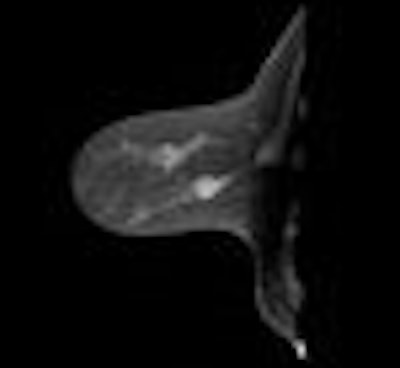
CT start-up firm Koning of West Henrietta, NY, will make its RSNA debut with a new work-in-progress CT scanner for breast and extremity use. The system is based on a conebeam x-ray tube and flat-panel digital detectors.
The company's first product, Koning CT for Breast and Extremities, uses a cone-shaped x-ray beam with a 30 x 40-cm flat-panel digital detector that captures images as a total volume rather than as individual slices. Koning believes that the configuration combines the advantages of digital projection imaging with CT, generating 3D images with true isotropic resolution and lower radiation dose levels than conventional CT scanners.

Koning believes that conebeam CT scanners have 10 times the data acquisition rates as conventional systems, with better contrast resolution and broad body resolution in a single rotation. The shorter scan times also result in fewer motion artifacts, and the scanners are less expensive, with half the annual ownership cost of conventional systems. Koning believes the system's technical specifications give it the performance equivalent to a 300-slice scanner.
Koning sees a future in which conebeam CT scanners are deployed in emergency rooms, intensive care units, image-guided surgical suites, and other locations where conventional CT systems cannot be used due to size, cost, or technical limitations. The Koning CT for Breast and Extremities scanner has not yet received marketing clearance in the U.S.
By Brian Casey
AuntMinnie.com staff writers
October 26, 2006
Copyright © 2006 AuntMinnie.com