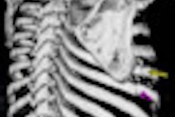
Quantifying hepatic fibrosis only with the texture pattern of the liver is an extremely difficult task for both radiologists and computers, leading to low diagnostic accuracy, said Xuejun Zhang, PhD, of Guangxi University.
While some researchers have been attempting to develop intelligent algorithms to stage fibrosis, their techniques have always worked on individual sequenced images or modalities. This makes it difficult to figure out the best approach to recommend to doctors, Zhang said.
In an attempt to discover the best approach, the group scanned several hundred patients in at least four different sequenced phases using different CT and MRI scanners.
"The accuracy rates outputted from different modalities, sequenced phased images, and texture features could not only tell us ... the optimal set of fibrotic pattern, guiding the radiologists to select an important view from numerous medical images, but also make clear the information of how many and what kind of texture features should a computer scientist use in their [computer-aided detection (CAD)] study," Zhang told AuntMinnie.com.
What conclusion did the researchers draw? Sit in on this Tuesday morning presentation to find out.