Type A Aortic Dissection:
The patient was a 47 year old female with a history of chronic renal insufficiency who presented to the emergency department with 11 out of 10 chest pain that radiated to her back. Her systolic blood pressure was elevated (160 mm Hg), but not severely.(Click on the small images to view the larger radiographs)
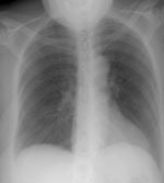
The chest radiograph was unremarkable. Clips in the lower neck were related to prior parathyroid surgery.

A chest CT was dome due to the severity of the patients symptoms. The
scan demonstrated an aortic dissection involving both the ascending and
descending aorta. The images of the chest revealed that the dissection
extended into the great vessels arising from the aortic arch (red arrows).

Images from the abdomen revealed the dissection extending into the superior
mesenteric artery with severe compromise of the native lumen (red arrow).


