Chronic Pulmonary Emboli:
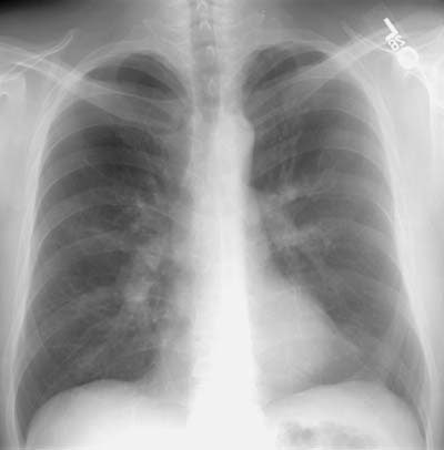
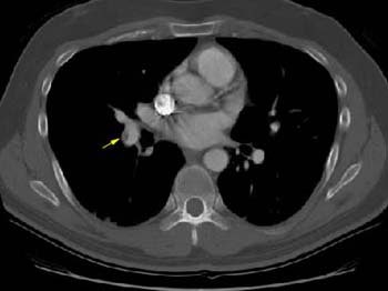
The patient presented for evaluation of progressive shortness of breath. The chest radiograph demonstrated prominent central pulmonary arteries. A cardiac echo suggested elevated pulmonary artery pressures indicative of pulmonary artery hypertension. A helical CT scan was performed and demonstrated the presence of arterial thrombus. Some vessels were completely occluded. An eccentric, mural thrombus could be seen in the right lower lobe pulmonary artery.(Click small images to view larger radiographs if desired)
CXR demonstrated prominent central vascular shadows:
CT scan revealed eccentric thrombus in the right lower lobe pulmonary artery:
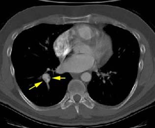
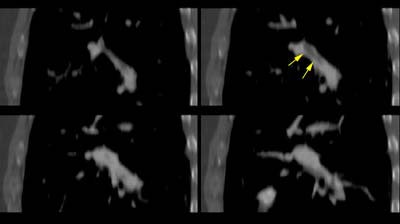
Helical CT permits reconstruction images along the course of the vessel. In this case the eccentric thrombus in the right lower lobe pulmonary artery is well seen:
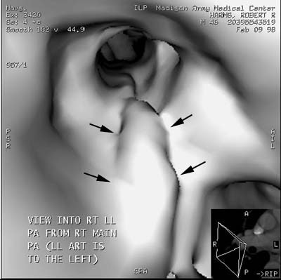
Endolumenal shaded surface images can also be performed:
