Pleural anatomy:
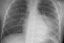
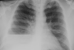
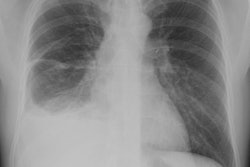
The visceral pleura covers the lungs and forms the interlobar fissures. The parietal pleura covers the mediastinum, diaphragm, and inner surface of the thorax. The normal thickness of the combined visceral and parietla pleura is only 0.2-0.4 mm and it is usually not visible radiographically. The parietal pleura is supplied by systemic capillary vessels arising from the intercostal arteries and drains into the right atrium via the azygous and internal mammary veins. The visceral pleura is supplied by pulmonary arterial capillaries (and to a lesser extent by the bronchial arteries) and drains mainly into the pulmonary veins.Lymphatic drainage of the visceral pleura is via the lymphatic plexus that covers the surface of the lung which drains cetrally along the bronchovascular bundles. There is normally only a thin film of fluid (2 to 20 ml) within the potential space between the visceral and parietal pleura. Normal lung fissures are less than 1 mm thick. Pleural fluid formation is thought to be governed by Starling's law of transcapillary exchange. There is a slight net movement of fluid into the pleural space from the parietal pleura, so that about 500 mL is created each day. The lymphatic drainage of the pleural space is through the parietal pleura- fluid leaves the pleural space via stomas in the parietal pleura which communicate with the chest wall lymphatics (intercostal, internal mammary, and mediastinal lymph node chains).Focal pleural disease is usually recognized as being extraparenchymal on CXR's by the presence of an "incomplete border" sign. Another finding characteristic of pleural disease is that the abnormality typically produces a smooth interface that has obtuse angles with the overlying aerated lung.
References:
(1) Radiographics 1997; 17: 63-79
(2) J Thorac Imaging 1998; Patz EF, et al. Percutaneous drainage of pleural collections. 13: 83-92.
(3) ACR Syllabus #40, p.142