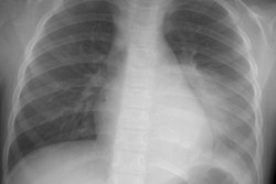
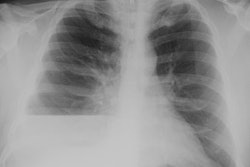
Hemothorax:
Clinical:
Hemothorax can occur secondary to trauma, bleeding diathesis, pulmonary infarct, catamenial hemorrhage, or infection. Blunt truama is a common cause of hemothorax. Massive hemothorax is defined as a hemothorax exceeding 1 liter with clinical signs of shock and hypoperfusion [1]. If the hemothorax occupies greater than one-half of the volume of the hemithorax, damage to a major vessel is likely, and thoracotomy is usually required for effective treatment (pleural hemorrhage in excess of 300 ml/hour is usually due to intercostal artery, hilar or mediastinal vessel injury. Bleeding from the lung is not usually this significant as passive atelectasis tends to tampnade the bleeding site. Complications of hemothorax include empyema (2-25% of patients), fibrothorax (fewer than 5% of patients), and pleural calcifications.X-ray:
On CT, a hemothorax usually appears hetergoeneous with a HU of 35-70 [1]. A fluid-hematocrit level can be seen. As the effusion begins to clot, loculations develop within the pleural fluid (fibrin balls).REFERENCES:
(1) (11) Radiographics 2008; Kaewlai R, et al. Multidetector CT of blunt thoracic trauma. 28: 1555-1570
(2) ACR Syllabus #40: p.464-465