J Thorac Imaging 1996;11(4):250-259
High-resolution CT findings of lung disease in patients with polymyositis and dermatomyositis.
Ikezoe J, Johkoh T, Kohno N, Takeuchi N, Ichikado K, Nakamura H
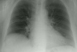
The purpose of this study was to determine the type and distribution of interstitial lung disease seen in patients with polymyositis and dermatomyositis, using high-resolution computed tomography (HRCT). The presence and distribution of high-resolution CT findings were retrospectively evaluated in 25 patients with polymyositis/dermatiomyositis. In 14 patients, a pathological diagnosis of pulmonary disease was obtained. Three lung specimens were also studied. Results showed that 23 patients had abnormal HRCT showing the following abnormalities: ground glass opacities (92%), linear opacities (92%), irregular interfaces (88%), airspace consolidation (52%), parenchymal micronodules (28%), and honeycombing (16%). A relatively high prevalence of airspace consolidation (52%) and a low prevalence of honeycombing (16%) were observed. Two patients with extensive consolidation proved to have diffuse alveolar damage; eight patients with either subpleural band-like opacities (n = 5) and/or airspace consolidation (n = 7) had bronchiolitis obliterans organizing pneumonia; four patients with honeycombing had usual interstitial pneumonitis. We conclude that HRCT findings in patients with polymyositis/dermatomyositis are nonspecific. However, a high prevalence of airspace consolidation and a low prevalence of honeycombing were observed. Predominant HRCT patterns are suggestive of the pathologic processes occurring in polymyositis/dermatomyositis.
PMID: 8892194, MUID: 97047274