Radiology 1994 Nov;193(2):375-382
Lung changes in rheumatoid arthritis: CT findings.
Remy-Jardin M, Remy J, Cortet B, Mauri F, Delcambre B
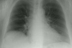
PURPOSE: To evaluate lung changes in rheumatoid arthritis (RA). MATERIALS AND METHODS: The authors reviewed the computed tomographic (CT) scans from 84 patients with RA with a mean articular disease duration (+/- standard deviation) of 12 years +/- 8 (range, 0.3-45 years). Fifteen patients underwent sequential CT evaluation during 5-65-month follow-up (mean, 18 months). RESULTS: Thirty-eight patients (49%) had abnormal CT scans showing the following abnormalities: (a) bronchiectasis and/or bronchiolectasis (n = 23, 30%), (b) pulmonary nodules (n = 17, 22%), (c) subpleural micronodules and/or pseudoplaques (n = 13, 17%), (d) nonseptal linear attenuation (n = 14, 18%), (e) areas of ground-glass attenuation (n = 11, 14%), and (f) honeycombing (n = 8, 10%). Abnormal CT examinations were recorded in 11 of 38 asymptomatic patients (29%) and 27 of 39 symptomatic patients (69%). The following CT abnormalities were found with a significantly higher frequency among patients with respiratory symptoms: (a) bronchiectasis and/or bronchiolectasis, (b) rounded areas of attenuation, (c) areas of ground-glass attenuation, and (d) honeycombing. CONCLUSION: CT may be a useful noninvasive tool for recognition of RA-associated lung disease with special emphasis on bronchial and bronchiolar changes.
PMID: 7972746, MUID: 95062972