Radiology 1995 Sep;196(3):835-840
Discrete lung involvement in systemic lupus erythematosus: CT assessment.
Bankier AA, Kiener HP, Wiesmayr MN, Fleischmann D, Kontrus M, Herold CJ, Graninger W, Hubsch P
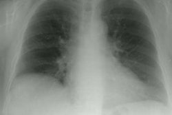
PURPOSE: To study the computed tomographic (CT) appearance of early lung involvement in systemic lupus erythematosus (SLE). MATERIALS AND METHODS: In a prospective study, 48 patients with serologically confirmed SLE but no prior clinical evidence of lung involvement underwent chest radiography, CT, and lung function tests. Radiographs and CT scans were compared, and CT scans were evaluated for signs suggestive of parenchymal and pleural disease. Extent and distribution of disease were determined. CT findings were correlated with clinical and functional data. RESULTS: Of 45 patients with normal chest radiographs, 17 (38%) had abnormal CT findings. Extent of disease was statistically significantly correlated with duration of clinical history (r = .93) and decreased single-breath diffusing capacity for carbon monoxide (r = .8) and ratio of forced expiratory volume in 1 second to forced vital capacity (r = .77). CONCLUSION: CT is superior to chest radiography for detection of functionally relevant pulmonary disease and is an important adjunct in early assessment of SLE.
PMID: 7644652, MUID: 95372540