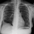
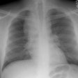
Sarcoid in a patient with a history of breast cancer:
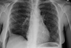
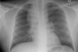
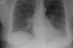
(Click on the small image to view the larger radiograph)The PA CXR revealed the patient to be status post a left mastectomy
for breast cancer. There are subtle, small nodular densities seen in the
right upper lobe.
A chest CT scan was done due to the suspicion for metastatic disease.
The exam revealed mutliple small nodules within the right upper lobe distributed
along the bronchovascular interstitium, the major fissure, and subpleurally.
These findings were associated with some evidence of distortion of the
underlying lung architexture. A few scattered nodules could be found in
the left upper lobe. The patient went to lung biopsy and the histologic
analysis revealed non-caseating granulomas consistent with sarcoidosis.
There was no evidence of metastatic disease.

