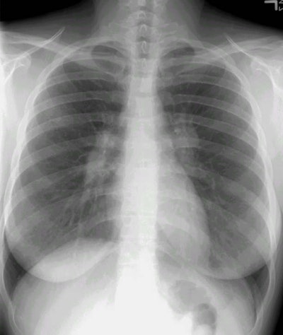
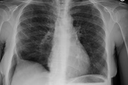
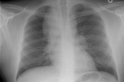
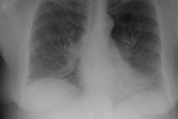
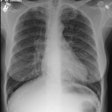
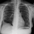
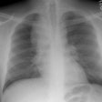
Sarcoid Stage I:
The patient was an asymptomatic active duty troop, who had the chest radiograph performed as a routine screening exam prior to ranger training. The PA chest radiograph demonstrates bilateral hilar fullness (compatible with adenopathy), a slight convexity to the aorto-pulmonary window region (consistent with mediastinal adenopathy), and subtle right paratracheal thickening (also suspicious for adenoapthy). (Click here if you would like to view the lateral exam).
A transbronchial biopsy confirmed the diagnosis of sarcoid. At our institution, asymptomatic patients do not undergo CT scanning. The patients are followed in pulmonary clinic with serial chest radiograph evaluation.
