CHICAGO - If radiology has a theme in 2001, it's that multiple modalities, working together, are sometimes better than one. The theory was recently applied to pancreatic cancer, perennially difficult to distinguish from chronic pancreatitis. And it turns out that combined MRI, MR cholangiopancreatography (MRCP) and contrast-enhanced dual-phase 3-D MR angiography can sometimes tell them apart.
In a presentation Sunday at the RSNA's pancreatic cancer scientific session, Dr. Ah Young Kim from the University of Ulsan in Seoul, South Korea presented her group's research aimed at determining the value of the three techniques in diagnosing and determining the resectability of pancreatic cancer.
"Despite rapid [progress] in MR imaging, it is still difficult to distinguish pancreatic cancer from chronic pancreatitis on the basis of known MR imaging criteria because both demonstrate low signal intensity," she said.
The 3-year study evaluated 126 consecutive patients (82 men and 44 women, ages 18-35), most with pathologic diagnosis based on biopsy or surgery.
MR imaging was performed with a 1.5-tesla scanner using breathhold techniques, including T1- and T2-weighted images with and without fat suppression.
MRCP was performed using both thick-slab RARE (rapid acquisition relaxation-enhanced), and multislice HASTE (half-Fourier fast spin-echo) techniques, as well as 3-D MRA using gadolinium.
Of the 126 patients, only the 93 patients who underwent surgical resection or open biopsy within 2 weeks of MR imaging were included in the study. Two readers blinded to the results retrospectively analyzed all of the MR images to assess vascular invasion, lymph node metastasis, and invasion to adjacent organs, Kim said.
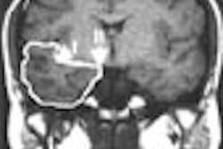
MRI correctly diagnosed 90/93 patients, with a sensitivity of 97% and specificity of 52%, Kim said. A pancreatic mass was present in 90/93 pancreatic cancers, and in 53 patients with chronic pancreatitis. Pseudocysts were found in 7 patients, and a calcified mass in 1 patient.
The mass contours were significantly smoother in most cases of chronic pancreatitis, and more ragged in most pancreatic carcinomas. In addition, the double duct sign is a statistically significant feature of pancreatic carcinoma, she said.
"Also, multiple sacular side branches were more frequent in pancreatitis," Kim said.
The overall diagnostic sensitivity and specificity of combined MR imaging were 93% and 85%, respectively, for vascular invasion, 64% and 84% for lymph node metastasis, 85% and 91% for duodenal invasion, 89% and 94% for hepatic metastases. MR missed 8 cases of omentum and duodenal seeding, however.
"The presence of the pancreatic mass, double duct sign, and irregular vasculature shown in combined MR imaging seems to be a valuable diagnostic finding for the evaluation of pancreatic cancer," Kim said.
By Eric BarnesAuntMinnie.com staff writer
November 26, 2001
For the rest of our coverage of the 2001 RSNA meeting, go to our RADCast@RSNA 2001.
Copyright © 2001 AuntMinnie.com