VIENNA - For a patient with acute or chronic hip pain whose x-ray results come up negative, Italian radiologists suggest using dynamic contrast-enhanced MRI instead.
Dr. Gabriella Cerone and colleagues from the Istituto di Radiologia dell’Universita in L’Aquila studied 20 patients with clinical evidence of hip pain and normal conventional x-ray exams. First, standard MRI was performed on a 1.5-tesla scanner with and without fat saturation.
After intravenous injection of Gd-DTPA contrast, additional MR images were acquired with spin-echo T1-weighted sequences on axial and coronal planes, with and without fat saturation.
In six cases, a quantitative analysis of femoral head signal intensity was performed before and after the contrast agent was administered. MRI follow-up was performed in 17 patients two months after the initial exam.
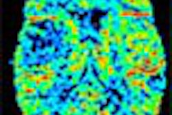
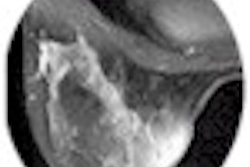
In all cases, the standard MRI showed anomalous signal intensity of the femoral head related to bone marrow edema syndrome (BMES). After the contrast medium was administered, a homogenous, diffuse enhancement with normalization of signal intensity was observed in 17 out of 20 cases.
"On the basal MRI, the deep difference of intensity values is evident between the two femoral heads. Whereas on the contrast-enhanced MRI, we see a normalization of the signal intensity because of the presence of active vascularization in the area," Cerone said.
At the two-month follow-up, all 17 patients had complete recovery of bone marrow edema after treatment, Cerone said. In the remaining three cases, the persistence of small areas of low signal intensity after contrast administration was related to the early stages of bone necrosis.
"We recommend intravenous paramagnetic MRI for the early differentiation of bone necrosis in these patients with negative plain film. [MRI] leads to early diagnosis, which has positive therapeutic value," she concluded.
The Italian study reiterated the findings of other recent studies. French radiologists found MRI best suited for pinpointing various hip disorders, including avascular bone necrosis of the femur head, articular effusions, and bone marrow edema (Journal de Radiologie, March 2000, Vol.81: Supplement 3, pp.392-408).
By Shalmali Pal
AuntMinnie.com staff writer
March 3, 2001
Click here to post your comments about this story. Please include the headline of the article in your message.
Copyright © 2001 AuntMinnie.com