While the exact mechanism of HIV dementia is unknown, pathological data have shown that central nervous system changes are common in HIV infection, even in the early stages of disease. Yet researchers have lacked a method of assessing CNS involvement in asymptomatic patients.
A recent study published in Magnetic Resonance Imaging found that MR spectroscopy allows for early detection of cerebral metabolite changes in neurologically asymptomatic HIV-infected patients with normal MRI results, providing the first reliable method of detecting early brain damage caused by HIV. In 30 asymptomatic seropositive patients, the authors found statistically significant decreases in two metabolite ratios compared to a control group.
Drs. Nitaya Suwanwela, Praphan Phanuphak, and others from the radiology department of the Chulalongkorn University Faculty of Medicine in Bangkok, Thailand, performed localized proton MR spectroscopy followed by MRI on 30 HIV-seropositive patients. Eighteen men and 12 women (ages 20-38) were included in the study. All of the patients were neurologically asymptomatic at various stages of HIV infection. None were taking antiretroviral medication, or had evidence of cerebral atrophy. Thirteen seronegative healthy volunteers comprised a control group.
"CNS involvement is a common feature of HIV infection," the authors wrote. "Early after systemic infection, HIV invades the cerebrospinal fluid space and also the brain parenchyma, presumably via infected monocytes/macrophages and T-cells that can traverse the blood-CSF and blood-brain barriers. During the early stages of infection, neither neurological abnormalities nor MRI changes could be found" (Magnetic Resonance Imaging, September 2000, Vol.18: 7, pp.859-865).
While previous studies have evaluated changes in brain metabolites of patients in the late stages of disease, the few that included early-stage asymptomatic patients found no clear diminution in metabolites, save for statistically insignificant trends toward reduced NAA/Cr and NAA/Cho ratios.
Thus, the researchers' goal was to determine if MR spectroscopy could locate any brain abnormalities in neurologically asymptomatic HIV patients that could not be seen with MRI, to correlate the findings with normal subjects, to correlate the spectroscopic ratios with patients' CD4 lymphocyte counts, and finally, to determine the MR spectrum of the control group.
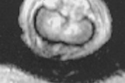
MRI was performed in all patients and control subjects using a 1.5-tesla scanner (Signa, GE Medical Systems, Waukesha, WI). The first pass was a sagittal T1-weighted sequence (repetition time [TR]/echo time [TE] = 2380/14, 84 msec, slice thickness 5.5 mm, gap 2.5 mm) followed by axial dual fast-spin-echo acquisition (repetition time [TR/echo time [TE] = 2380/14). The MR images were reviewed independently by two neuroradiologists blinded to clinical, immunologic, and spectroscopic results.
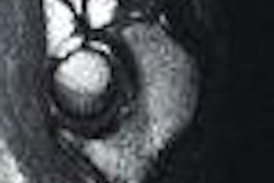
Localized proton spectroscopy was performed with automated single-voxel proton spectroscopy (PROBE/SV, GE Medical Systems, stimulated echo-pulse sequence, probe-S), using 2.5-KHz spectral width, 2,048 sample points, 128 averages [42], TE of 30 ms and TR of 2020 ms, the authors wrote.
The investigators used an 8-cc volume of interest, and voxels were placed in three brain regions, two in the centrum semiovale to evaluate subcortical white matter, and one in the thalmus to assess the deep gray matter.
They measured the four peeks of N-Acetylaspartate (NAA) at 2.02 ppm; creatine (Cr) at 3.02 ppm, choline (Cho) at 3.22 ppm and myoinositol (mI). The metabolite ratios of NAA/Cr, Cho/Cr, NAA/Cho and mI/Cho were then calculated for each case using the GE Sage spectral analysis package. The results were analyzed to evaluate significant differences in brain metabolite ratios in the centrum semiovale and thalmus.
In metabolite ratios, the authors found a statistically significant reduction in NAA/Cr and NAA/Cho in both the centrum semiovale (p<0.005) and thalamic areas (p<0.05) between the HIV-seropositive group and the control subjects. No statistically significant difference was found in Cho/Cr or mI/Cr in either region.
"Reduction of NAA/Cr and NAA/Cho ratios measured by MRS generally indicates neuronal damage or loss," they wrote. "In our study, significantly decreased ratios were found despite a normal MR imaging. We therefore conclude that the reduction of NAA might represent neuronal dysfunction or early neuronal damage without gross anatomical abnormalities of the brain, which may signify latent encephalopathic changes undetectable by MR imaging or routine neurological examination."
The Cho/Cr ratio is believed to be a crude measure of cell membrane material, so increased choline may indicate cellular injury or increased glial proliferation, according to the authors. In contrast to previous studies of late-stage HIV patients, they found no significant difference between their patients and the control group.
"Therefore, the Cho/Cr ratio may not be as sensitive as the NAA/Cr and NAA/Cho ratios to detect early changes in the brain of HIV patients," they wrote.
The researchers noted that increased mI is generally considered to be evidence of glial proliferation. Increased levels of this metabolite have been found in Alzheimer's patients, and in patients with advanced AIDS dementia complex. However, the authors found no significant difference between the patients and the control group.
"Gliosis is apparently not a major component of early brain dysfunction in neurologically asymptomatic HIV patients," they wrote.
Finally, the authors found that metabolite ratio changes of NAA/Cr and NAA/Cho were more pronounced in the white matter (centrum semiovale) than in the deep gray matter (thalamus).
"This finding correlates well with the pathological study showing early involvement of the disease in the white matter," they wrote. "The subcortical gray was affected at a lower frequency, followed by the cerebral cortex."
As for immune status, linear regression analysis of the data found a slight negative trend toward correlation between CD4 counts and NAA/Cr ratios in patients with advanced disease, but no statistically significant correlation between Cho/Cr or mI/Cr ratios.
"To date, this is the largest case-control study of metabolite ratios measured by MRS in neurologically asymptomatic HIV patients who had normal MR imagings.... Proton magnetic resonance spectroscopy is more sensitive than MR imaging for evaluation of CNS involvement in HIV infection," they concluded.
By Eric BarnesAuntMinnie.com staff writer
November 22, 2000
Click here to post your comments about this story.
Copyright © 2000 AuntMinnie.com