Thanks to a noise-reduction algorithm known as adaptive statistical iterative reconstruction (ASIR), virtual colonoscopy scans acquired at half the normal dose delivered image quality equivalent to standard images, according to a study published in the July issue of the American Journal of Roentgenology.
Researchers from the Mayo Clinic in Scottsdale, AZ, found no significant differences in image quality or noise between standard-dose images and those acquired at half the dose using ASIR.
ASIR is a proprietary algorithm that uses an iterative reconstruction technique to reduce dose without reducing image quality compared to standard filtered back projection (FBP) images, the authors wrote.
"Unlike FBR, ASIR does not assume noise is evenly distributed across the entire image, but instead uses a mathematic model to identify and remove individual projections that are noisy and deviate from the model," explained lead investigator Kristina Flicek, MD, and colleagues (AJR, July 2010, Vol. 195:7, pp. 126-131).
Although virtual colonoscopy (also known as CT colonography or CTC) is intended to be noisier and typically acquired at doses of 2 mSv or less, patients need assurance that the dose they receive is low as possible. Moreover, because CTC is often repeated during the patient's lifetime in a subsequent screening or follow-up scan, physicians have an interest in exploring the lower limits of radiation doses that will still deliver high sensitivity and specificity.
"Despite the lack of scientific data measuring future cancer risk from radiation exposure, patients remain concerned about radiation exposure," the authors wrote. "In response to public concern and the desire for the best patient care, we sought to reduce the radiation dose during CT whenever possible without compromising image quality."
The study was divided into two parts: an initial phantom study to determine the dose and ASIR level for the patient arm of the study, followed by the patient study that used the supine image acquisition (using standard dose, 50 mAs) and the prone position with a 50% dose reduction (25 mAs) and the addition of the proprietary ASIR noise-reduction algorithm (GE Healthcare, Chalfont St. Giles, U.K.) set at 40%.
For the phantom arm of the study, a Silastic air-filled phantom was imaged on a 64-detector-row scanner (Discovery CT750HD, GE) at 120 kVp and 1.25-mm collimation, with a 0.625-mm slice thickness.
The phantom was scanned at 50 mAs without ASIR and at several mAs settings (40, 30, 20, and 10 mAs) both with and without the application. For each mAs setting, image data were reconstructed using 0%, 20%, 40%, 60%, 80%, and 100% weighting of ASIR, which is typically blended with the native image data to achieve the optimal combination of noise reduction and image quality. Noise was assessed, and overall image quality was rated on a scale of 0 (nondiagnostic) to 4 (highest image quality) by an experienced gastrointestinal radiologist.
In the patient arm of the study, 18 patients (age, 35-84 years; mean, 68) underwent standard cathartic bowel cleansing followed by mechanical insufflation of the colon. The patients were scanned in the supine position and at a reduced dose of 25 mAs with 40% ASIR in the prone position, using the same scanner as in the phantom study.
Three experienced radiologists blinded to the scanning techniques assessed 2D and 3D image quality and noise at three different colon locations. A score difference of ≥ 1 was considered clinically important. Actual noise measures were compared between the standard-dose and low-dose acquisitions, the authors wrote.
The phantom study showed image noise reduction that correlated with a higher percentage of ASIR. Image noise decreased as the percentage of ASIR was increased and as the mAs was increased.
For example, the dose-length product (DLP) for a patient with a body mass index (BMI) of less than 20 was 183 for routine CTC acquisition and 97.7 for the ASIR technique. For a patient with a BMI of 30 or greater, DLP was 303.4 for the routine acquisition and 163.4 for the low-dose ASIR scan. The overall DLP of 230.8 for the standard technique dropped to 121.2 with the use of ASIR.
"At the standard dose of 50 mAs without ASIR, noise was measured at 12.7 HU," Flicek and colleagues wrote. And by varying the ASIR amounts, the noise levels at lower doses could match or exceed this baseline value.
"For example, an exposure of 30 mAs at 60% ASIR was associated with a noise level of 10.7 HU," they wrote. Even when dialing the exposure down to 10 mAs with progressively higher levels of ASIR, noise measurements were equal to or lower than standard-dose noise measurement without ASIR.
Ratings of 2D image quality reached maximum image quality (4) for all dose levels when ASIR was applied anywhere between 20% and 100%, they wrote. For images acquired at 50 mAs without ASIR, the baseline rating for 3D image quality was 3.
Low image quality scores were more dependent on dose settings than on the use of ASIR, while higher-quality images were improved with the addition of ASIR.
The researchers found no differences of one point or more in the comparison of subjective image noise and image quality scores between routine and low-dose images.
Similarly, in the patient arm of the study, no significant image quality differences were seen between low- and standard-dose images at 40% ASIR, they wrote. Overall image quality was reduced for both image sets as body mass index increased. Measured image noise was less with the low-dose technique using ASIR.
 |
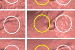
| Axial CTC images for comparison of standard versus low-dose ASIR technique at the level of splenic flexure. Above (A-C), 2D axial images -- supine image using standard-dose CTC with 50 mAs and 0% ASIR (A), prone image using 25 mAs and 0% ASIR (B), and prone image using 25 mAs and 40% ASIR (C) -- show ASIR reduces noise artifact to produce improved image. Below (D-G), 3D axial images without and with ASIR -- 50 mAs without ASIR (D), 50 mAs with ASIR (E), 25 mAs without ASIR (F), and 25 mAs with ASIR (G) -- show addition of ASIR reduces image artifact (mucosal nodularity and endoluminal floaters) with both dose settings. Images D and G are comparable despite reduced dose (2.1 versus 4.2 volume CT dose index). Images republished with permission of the American Roentgen Ray Society. |
 |
Image noise varied from 28.3 to 61.8 HU among the three patient size groups using standard CTC dose settings in the supine position. With the use of 50% dose reduction (25 mAs) and 40% ASIR, the mean noise measurement was lower, ranging from 23.7 to 51.2 HU. Objective noise increased with increases in patient size.
In both standard- and low-dose images, subjective image noise and image quality scores worsened as patient size increased. The standard-dose noise scores ranged from 2.9 to 3.8 HU in the smallest patients and from 2 to 2.8 in the largest. Low-dose noise scores ranged from 2.4 to 3.3 HU in the smallest patients and from 1.9 to 2.5 HU in the largest patients, the group reported.
"Our hypothesis, that the standard radiation dose for CTC (i.e., 50 mAs) could be reduced
50% when combined with ASIR without substantially affecting image quality, was validated both in a phantom and in humans," Flicek and colleagues wrote. "Objective image noise measures were actually lower at 25 mAs with ASIR than at a routine dose of 50 mAs without ASIR for all patients regardless of BMI."
The best subjective scores of image quality and image noise were found in the smallest patients, and both increased with patient size.
"Interestingly, however, for both routine- and low-dose images, image sharpness was graded slightly worse in smaller patients than average or large patients," the group noted. "This may be explained by less periaortic fat in the smaller patients so that the contrast between the unenhanced aorta and surrounding structures is less apparent, an issue exacerbated by lower radiation dose," they wrote.
Based on the encouraging results of the pilot study, the authors recommended further clinical evaluation in a larger patient cohort.
By Eric Barnes
AuntMinnie.com staff writer
June 21, 2010
Related Reading
MBIR aims to outshine ASIR for sharpness, CT dose reduction, May 18, 2010
Filtering software boosts CT image quality, lowers dose, May 7, 2010
Reduced dose can still yield acceptable pediatric CT images, April 22, 2010
Automated exposure control delivers uneven CT dose reduction, April 15, 2010
ASIR cuts dose in Crohn's disease patients, December 4, 2009
Copyright © 2010 AuntMinnie.com