SEATTLE - Fluorodeoxyglucose (FDG) PET has proved to be indispensable for evaluating primary musculoskeletal lesions, according to radiologist Dr. Frieda Feldman from Columbia University and Columbia Presbyterian Medical Center in New York City. In a presentation Tuesday at the American Roentgen Ray Society meeting, Feldman said that, in addition to its value in documenting and staging primary musculoskeletal lesions, PET improves accuracy by directing biopsy and evaluating both treatment response and recurrence.
"FDG is the current favorite, but it's really far from current," Feldman said. "Its initial introduction was by [M.] Blau in 1962, and it fast became the basic bone-scanning agent for a whole decade, until it was replaced by a single photon-emitting diphosphinate. But it bounced back in the '80s for two very good reasons. First, the increasingly sophisticated data analysis of scans ... and because it's a fooler, it use the [body's] glucose transport method to enter the cell, where it becomes phosphorylated by hexokinase, and loses its diffusability. It becomes trapped in the cell, and serves as a marker for glucose-avid malignancies."
The study looked at 20 patients with varied benign or malignant primary bone or soft-tissue lesions that were evaluated with FDG-PET, both before and after surgical intervention. FDG-PET scans were evaluated both quantitatively and qualitatively, and compared to contralateral regions and standard uptake values. The results were then correlated with radiographic, MRI, intraoperative and histologic findings.
Highlighting several cases, Feldman emphasized PET's potential in reconciling clinically discrepant imaging and histologic findings, especially in the problematic area of cartilage lesions.
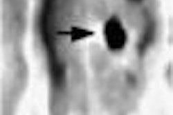
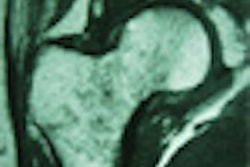
An elderly female patient with nondescript bone pain in the knee area and inconclusive MRI findings underwent open focal biopsy that resulted in a diagnosis of enchondroma. Subsequent PET-guided open biopsy changed the diagnosis to grade-1 chondrosarcoma.
Another patient had a similar dilemma, Feldman said. "Calcified cartilage-appearing lesions on an 80-year-old women, [only] larger and more painful, with a definitely positive bone scan. We went straight to [FDG]-PET, which showed a totally nonaggressive lesion with no metabolically active focus, with a contralateral axillary inflammatory node, but whose whole-body scan was completely negative, including the lung."
In addition to detecting and ruling out metastases, PET also serves as an intraoperative guide, particularly in sampling large and heterogeneous lesions, Feldman said.
In a 79-year-old patient with a heavily calcified lesion presumed to be juxtacortical chondroma, PET ruled out metastases and enabled the limb to be salvaged.
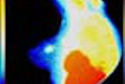
"Most malignancies exhibit generally increased FDG concentration, usually connoting a high-grade lesion," Feldman said. "However, malignant lesions that are more mature, or with heavy mineralization or necroses, may show centrally decreased FDG concentration, simulating what I like to call a dome. Conversely, depending on perfusion, there's a caveat. FDG uptake may be relatively diminished in slow-growing, well-differentiated, less-aggressive lesions."
Finally, in an 11-year-old girl, an initial attempt at limb salvage for osteosarcoma was deemed successful. However, FDG-PET was performed due to an increase in pain, which could have been due to any number of factors, such as loosening, infection, or prosthetic failure, Feldman said. Unfortunately, FDG-PET showed that the cancer had spread to the tibia, the lungs, and the proximal right humerus of the young patient.
In summary, PET's advantages include staging known tumors and detecting unknown metastases, Feldman said. It also shows promise in gauging aggressiveness, and improves accuracy through targeted biopsy, particularly in large heterogeneous tumors, thereby aiding treatment planning and the evaluation of treatment response and prognosis.
Moreover, PET is cost-effective because it allows the assessment of disease character, metabolism, and extent in a single scan, and reduces the complexity of post-treatment workups and their associated morbidity, she said.
"But the best is yet to come," Feldman said, "due to improving quantitative analytic techniques, and targeting different metabolic functions from the same tumor, with different tracers, including perfusion rates, for which oxygen-15[-labeled carbon dioxide] has been used, as well as technetium 99-m and c-11 methionine for protein synthesis, as well as glucose metabolism, thereby enabling targeted treatment of specific abnormal functions with tailor-made pharmaceuticals."
Finally, she said, PET's future lies in image fusion with MRI and CT, "which will make for one-stop testing and analysis, and hopefully, a more accurate and efficacious approach to visualization."
By Eric Barnes
AuntMinnie.com staff writer
May 2, 2001
Click here to post your comments about this story. Please include the headline of the article in your message.
Copyright © 2001 AuntMinnie.com