By combining partial-scan reconstruction and multislice spiral weighting, German researchers have devised a dedicated algorithm for electrocardiographically gated (ECG) multislice CT that covers cardiac volume in one breath-hold. They tested their method on 10 patients with varying degrees of arrhythmia and found that the technique offered high spatial resolution and good overall image quality.
Bernd Ohnesorge from Siemens Medical Engineering in Forchheim headed the multi-institutional team that included radiologists from the University of Munich and the University of Erlangen-Nurenberg. The group will update their results in a scientific session (974) at the RSNA conference later this month.
"In our study, we wanted to evaluate the suitability of the different ECG-gating techniques for generation of images in the diastolic and systolic phases of the cardiac cycle. We investigated this application for acquiring ECG-synchronized multisection spiral scans with heart rate-dependent table feed (pitch) adaptation," they wrote in the November issue of Radiology (Vol.217:2, pp.564-571).
The algorithm allows for the reconstruction of overlapping images at arbitrary z positions and during any given cardiac phase, the authors wrote. Each image must be reconstructed using a multislice, partial-scan data segment with an arbitrary temporal relation to the R wave of the ECG trace.
"Continuous volume coverage can be achieved only when the spiral pitch is adapted to the heart rate to avoid gaps between image stacks that are reconstructed by using data from different cardiac cycles. To achieve full volume coverage, the image stacks reconstructed in subsequent cardiac cycles must cover all z positions," they explained.
The 10 patients underwent spiral CT scans with a Somatom VolumeZoom (Siemens Medical Engineering, Forchheim, Germany) with simultaneous acquisition of four sections. Iopromide (Ultravist 300, Schering, Berlin) was injected intravenously at a flow rate of 3 mL/sec. Their ECG signals were recorded during multisection spiral scanning in order to match the spiral acquisition to specific phases of the cardiac cycle.
One of three phase-selection strategies was used:
- Relative-delay, in which the delay time is determined individually for each cardiac cycle as a given percentage of the R-R interval.
- Absolute-delay, where the start point of the reconstruction data interval is determined by fixed delay times prior to the onset of the R wave.
- Absolute-reverse, in which fixed times prior to the onset of the next R wave defines the start point of the reconstruction data interval.
The multislice data acquisition was obtained using a 500-msec full rotation time, which resulted in 250-msec temporal resolution and 4 x 1 mm, or 4 x 2.5 mm, collimated sections. Images were then reconstructed at 1-mm or 0.5-mm increments. Two observers examined the scans, assessing for calcified lesions and whether the images were free of motion artifacts in the diastolic cardiac phase.
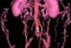
In three patients with considerable arrhythmia (55-82 beats per minute, 72-85 beats per minute, 110-124 beats per minute), the coronary arteries, including vessel calcifications, were clearly seen, the authors noted. The best results without motion artifacts were obtained using absolute-reverse ECG gating with a reconstruction interval of 450 msec for moderate heart rates and 350 msec for higher heart rates.
"In seven patients, cardiac anatomy, including the major coronary arterial branches, was depicted free of motion artifacts with an appropriate selection of the ECG-gating phase parameters. With 1.25-mm sections, smaller side branches could also be assessed reliably," they wrote.
The team also reported an improvement in volume reconstruction in the diastolic phase by using absolute-reverse ECG gating and a gain in z resolution with the use of 1.25-mm sections.
"In contrast to sequential CT scans, the z resolution of ECG-gated spiral images with 3-mm sections can be improved by using overlapping reconstruction with 1-mm section increments," they wrote. "Moreover, the fast scanning speed allows coverage of the entire heart with 1.25-mm sections within a single breath-hold."
Future research will use the technique for noninvasive screening of coronary arterial disease, comparing it with other noninvasive and invasive modalities, the authors concluded.
By Shalmali PalAuntMinnie.com staff writer
November 14, 2000
Click here to post your comments about this story.
Copyright © 2000 AuntMinnie.com