Shear-wave ultrasound elastography is a useful tool that may be deployed in many different organs, including the breast, prostate, thyroid, pancreas, and musculoskeletal system. But care must be taken to use the right protocols and to avoid artifacts and other pitfalls.
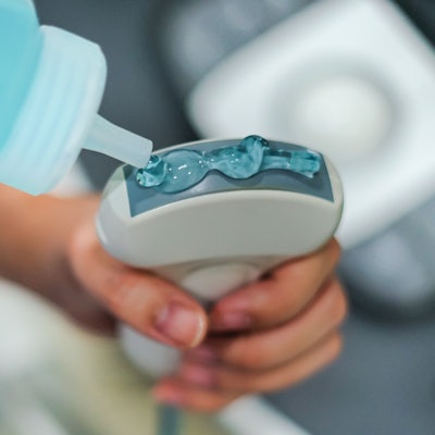
Elastography emerged in the 1990s, evolving from the classic physician's technique of using manual palpation to detect tissue stiffness, which can give clues to pathology. With ultrasound elastography, the stiffness of tissue is measured while under external pressure.
There are different types of ultrasound elastography. With strain elastography, a transducer or acoustic radiation force imaging (ARFI) may be used to apply stress on the tissue of interest and to assess stiffness in a field of view.
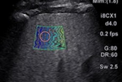
With shear-wave elastography, a newer method for measuring elasticity, an ARFI pulse generates shear waves in a region of interest in a process called point shear-wave elastography (p-SWE) -- or over a larger field of view using 2D or 3D techniques. The speed of the shear wave -- as measured in meters per second (m/s) or kilopascals (kPa) -- indicates stiffness.
 Dr. Giovanna Ferraioli.
"The shear-wave speed (SWS) is almost one thousand times lower than the velocity of [ultrasound] in tissues, the shear waves attenuate very rapidly, and some do not propagate in the simple fluids," explained Dr. Giovanna Ferraioli, a researcher at the Medical School University of Pavia in Italy, and colleagues in two related articles on how to perform shear-wave elastography recently published online in Medical Ultrasonography.
Ferraioli et al provided detailed guidance on how to perform shear-wave elastography by organ type. Below are some highlights from their advice.
Breast
In the breast, shear-wave elastography is useful for differentiating benign from malignant lesions, as malignant tissue tends to be stiffer. It is also playing a role in monitoring effects of neoadjuvant chemotherapy, Ferraoli et al noted.
The authors advised using a standard 5-cm-wide linear transducer for evaluating the breast. Higher frequencies should be used for evaluating superficial lesions, whereas lower frequencies are appropriate for lesions located deep in the breast.
Due to artifacts, there is a risk for false negatives with shear-wave ultrasound in the breast, but this can be minimized by using a color-coded "quality map" to evaluate shear-wave propagation and image quality, they advised. Combining strain elastography with shear-wave elastography can also help minimize artifacts that result in false-negative results.
Users should also be mindful of precompression -- if they apply too much, a lesion will appear stiffer than it actually is on elastography images.
"Therefore, the recommendation is to apply a large amount of gel and then place the [ultrasound] transducer on the breast," Ferraioli et al wrote. "The subcutaneous fatty tissue should appear in dark blue. If it appears in green or even red [that means] too much pre-compression is used and needs to be corrected."
Prostate
Prostate cancer lesions are most commonly found in the peripheral zone, which contains most prostatic glandular tissue. Shear-wave elastography has a high negative predictive value for evaluating prostate cancer lesions located in this zone and can be helpful for monitoring men with rising prostate-specific antigen (PSA) levels, potentially helping to avoid unnecessary biopsies, Ferraioli et al advised.
However, on shear-wave elastography, benign prostatic hypertrophy is stiff and the technique's utility is limited in other parts of the prostate gland, they wrote.
Shear-wave elastography of the prostate should be performed after a complete transrectal ultrasound study, Ferraioli et al advised. An endocavity transducer, which has shear-wave capabilities, should be used. Two-dimensional shear-wave elastography (2D-SWE) is needed for localizing areas of stiffness and for guiding biopsies.
"With p-SWE it is not possible to identify the area(s) of concern in a timely fashion or accurately," the authors explained.
The researchers advised holding the transducer lightly and centering the area of concern in the middle of the image. Precompression should be avoided and elastography images should be compared with B-mode images to avoid mistaking prostate calcifications, which have higher stiffness values, for malignancies.
Thyroid
Shear-wave elastography can step in as a noninvasive tool for evaluating thyroid nodules, which are very common, acting as an alternative for fine-needle aspiration (FNA) biopsy in some patients. A high-end linear transducer is needed for imaging, the authors advised. With p-SWE, users should acquire from three to 10 measurements, and with 2D-SWE, they should acquire at least three.
"An abundant quantity of gel should be used to avoid pre-compression, since it may alter tissue elastic modulus, thus causing artifacts," Ferraioli et al advised. "The operator places the transducer perpendicular to the target nodule without pressure, maintaining only slight contact with the skin."
It may also be challenging to image patients with prior neck surgery and its resulting fibrosis, the authors cautioned.
Musculoskeletal system
Ultrasound is a workhorse modality for imaging the musculoskeletal system, enabling dynamic visualization of muscles, tendons, and joints, and shear-wave techniques add to the breadth of information with an assessment of elastic properties.
"Additional evaluation of tissue elastic properties is a new technique that allows for the detection and confirmation of pathologies such as neoplastic masses, and nonneoplastic changes such as tendinitis, myositis, fasciopathy, sprains and tears," Ferraioli and colleagues noted.
A high-frequency linear probe is typically used for imaging the musculoskeletal system, though a lower-frequency linear transducer or a convex transducer may be more appropriate for structures in deeper locations, the authors suggested.
Users should apply mild pressure with the transducer of choice and place the transducer longitudinally to the muscle fibers, Ferraioli et al advised.
The authors explained parameters for both p-SWE and 2D-SWE of the musculoskeletal system.
"With real-time 2D-SWE, stabilization of the image for a few seconds before the measurement is taken is very important," the authors wrote. "As soft tissues give a wide range of stiffness, it is recommended to adjust the scale to maximum. SWE has a high specificity in the detection of tendinopathy and subclinical tendinopathy, showing the softening of the tissues."
Users should avoid precompression because this could increase the appearance of stiffness in tissues, the authors advised.
Lymph nodes
Ultrasound elastography has become a useful technique for differentiating benign from abnormal conditions in the lymph nodes. Malignant conditions like metastatic lymphadenopathy and lymphoma are associated with greater stiffness than benign findings, such as areas of infection or inflammation, Ferraioli et al wrote.
In cervical lymph nodes, shear-wave elastography has a reported sensitivity of 81% and specificity of 85%, the authors noted.
Imagers must know what normal lymph nodes look like on B-mode ultrasound and should use a linear probe with a wide range of frequencies, Ferraioli and colleagues advised. As with other applications, lower frequencies should be used for areas of interest in deeper locations.
To minimize artifacts, patients should avoid swallowing and should hold their breath during imaging.
"During the elastographic examination, the absence of a result indicates a poor quality of the study," Ferraioli et al advised. "More than one measurement must be performed to improve the quality of the study."
Pancreas
As with other organ types, malignancies in the pancreas are stiffer than benign areas, such as healthy parenchyma. In the pancreas, shear-wave ultrasonography is helpful for distinguishing benign from malignant lesions, guiding biopsies, and characterizing stiffness in suspected chronic pancreatitis, Ferraioli et al wrote.
However, the authors added, "Due to its limited clinical role in the differential diagnosis, elastography cannot replace pathological examination with FNA or biopsy."
The authors suggested using 2D-SWE for quantitative and qualitative results and to select a convex probe.
"To completely visualize the pancreas, it is sometimes useful to examine the patient during an inspiration or expiration," Ferraioli et al said. "Compression with the transducer to displace bowel gas may allow [visualization of] the retroperitoneum and [improved visualization of] the pancreatic gland."
Malignant lesions vary in the degree of stiffness and need to be completely evaluated, the authors noted.
"Regarding the methodology, more than one elastogram and more than one measurement must always be performed," Ferraioli et al wrote. "It is reported that five measurements need to be performed for each lesion or area under investigation."
Liver
Finally, shear-wave elastography has become particularly valuable for applications in the liver, such as hepatocellular carcinoma, chronic liver disease, and assessing the severity of liver fibrosis.