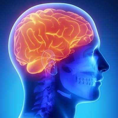
CT shows that COVID-19 patients who require oxygen treatment and those who have fever have reduced gray-matter volume in the frontal-temporal lobe of the brain months after hospital discharge, according to a study published May 11 in Neurobiology of Stress.
The study underscores the long-term negative effects of the illness, researchers from the Georgia Institute of Technology in Atlanta said in a statement.
"The study found lower gray-matter volume in this brain region was associated with a higher level of disability among COVID-19 patients, even six months after hospital discharge," the institute said.
The brain's gray matter is crucial for information processing, and damage to it can affect how neurons communicate, wrote a team led by graduate student Kuaikuai Duan. And although researchers have begun to document neurological complications in COVID-19 patients, how the disease affects the brain needs clarification.
"Science has shown that the brain's structure affects its function, and abnormal brain imaging has emerged as a major feature of COVID-19," Duan said in the institute's statement. "Previous studies have examined how the brain is affected by COVID-19 using a univariate approach, but ours is the first to use a multivariate, data-driven approach to link these changes to specific COVID-19 characteristics (for example, fever and lack of oxygen) and outcome (disability level)."
Duan and colleagues conducted a study to assess COVID-19's effect on gray matter, analyzing CT scans from 120 patients, 58 who had acute COVID-19 and 62 who did not. The researchers used source-based morphometry analysis to evaluate the data. Patients' disability levels were assessed with the six-point modified Rankin scale, at preadmission, discharge, and six-month follow-up; higher numbers indicated more severe disability.
The investigators found that people with higher levels of disability at discharge from the hospital and six months later had reduced gray-matter volume in the superior, medial, and middle frontal gyri, even when the group controlled for cerebrovascular diseases. Patients who needed oxygen and who had fever during the course of their illness also had reduced gray-matter volume.
Duan's team noted reduced gray matter in patients who had experienced agitation during their illness compared with those who had not -- suggesting that "gray-matter changes in the frontal region of the brain may underlie the mood disturbances commonly exhibited by COVID-19 patients."
The results suggest a new way to predict COVID-19 patient outcomes, according to the authors.
"[Our study implies] that the frontal-temporal network could potentially be employed as a biomarker for prognosis and evaluation of treatment of COVID-19," they concluded.