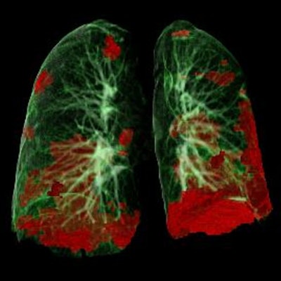
CT images of severe COVID-19 may help clinicians predict patients' vulnerability to neurological conditions, and thus abnormal findings on MRI, according to a study by University of Cincinnati researchers published March 12 in the American Journal of Neuroradiology.
The findings confirm imaging's key role in the COVID-19 pandemic, said lead author Dr. Abdelkader Mahammedi of the University of Cincinnati Gardner Neuroscience Institute in Ohio, in a statement released by the institution.
Mahammedi and colleagues conducted a study that included data from 135 patients hospitalized with COVID-19 between March and June 2020 at healthcare facilities in Spain, Italy, and Brazil as well as at the University of Cincinnati. Patients experienced both lung and brain effects from the disease and had both CT and MRI images available.
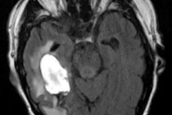
Of the total number of patients with abnormal lung scans and neurological symptoms, 36% had abnormal findings on brain imaging and were more likely to experience symptoms of stroke, the group found.
The study results should help physicians better identify patients more likely to develop brain imaging abnormalities based on the severity of disease found on their CT scans, according to Mahammedi.