SAN FRANCISCO - FDG-PET can pinpoint distant esophageal metastases and potentially increase long-term survival after induction therapy, but whether it can alter the course of cancer treatment has yet to be determined. These are the conclusions reached by two groups of researchers at the American Society of Clinical Oncology meeting on Monday.
Dr. Robert Downey and his colleagues from Memorial Sloan-Kettering Cancer Center in New York City prospectively evaluated 18fluorodeoxyglucose (FDG) PET for measuring the pathologic response of esophageal cancer to a combination of chemotherapy and radiation therapy, also known as induction therapy.
"PET has been proven valuable in the staging of patients with untreated esophageal cancer," Downey said. "The metabolism of both normal and malignant tissues is significantly altered by anti-neoplastic therapies. The malignancy can have a reduced signal both because of cell death and lowered metabolism. The surrounding normal tissues can have increased FDG uptake because of inflammation. The overall effects of these factors on the accuracy of PET after induction therapy is not known."
Forty patients with biopsy-proven esophageal cancer were enrolled in the study, which took place between 1997 and 1999. Imaging was done pre-treatment with the GE Advance PET scanner (GE Medical Systems, Waukesha, WI) and in conjunction with standard imaging studies, such as CT or endoscopic ultrasound. Following induction therapy, restaging was performed with FDG-PET, and followed by surgical resection where appropriate.
"All PET scans were performed on a dedicated scanner and encompassed at least the neck, chest, abdomen, and pelvis. Patients fasted for 6 hours prior to the injection of more than 10 mCi of FDG. Post-emission and transmission scans were performed on all patients, allowing a filtered back projection algorithm to be used for reconstruction," Downey said.
One nuclear medicine specialist and one radiologist reviewed the images. A pathologist reviewed all resected primary tumors and assigned a percentage treatment effect. A thoracic surgeon assigned a final pathological stage based on all available clinical materials.
According to the results, pre-treatment FDG-PET scans revealed inoperable metastatic disease in 20% of the patients, and a second primary malignancy in 2%. Of the 32 patients with clinically operable esophageal cancer, 27 received pre-operative chemotherapy and radiation therapy (5 patients withdrew from the study).
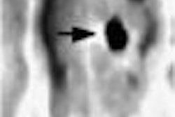
FDG-PET restaging was performed in 23 of these patients. The standardized uptake value (SUV) of the primary tumor decreased by a median of 53% in these patients between the two PET studies.
In addition, "patients who have a less than 57% increase in SUV had a median disease-free survival of 17 months," Downey said. "The changes in FDG-PET before and after induction therapy may correlate with disease-free survival."
While the study confirms the benefits of PET for the initial staging of metastatic disease, the modality fell short in one respect. PET studies that are performed soon after induction therapy (in this case, 2 weeks) did not appear useful for the detection of new sites or locoregional sites of disease, Downey concluded.
In a second paper, Dr. Patrick Flamen from the University Hospital in Leuven, Belgium discussed the advantages of whole-body FDG-PET to predict the pathologic response of esophageal cancer after preoperative chemoradiation therapy (CRT).
Because the resection success rate of patients is difficult to determine in these patients, many undergo CRT in order to downstage the tumor and increase resectability, Flamen said. However, only 30-50% of these patients who undergo this treatment protocol will enjoy overall long-term survival.
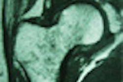
‘There is a strong need for imaging tools that enable the prediction of response rate and also allow for the differentiation of responders and non-responders after treatment," he said. "Conventional imaging modalities such as CT, MR, and endoscopic ultrasound are not very effective for evaluating response, because these instruments provide structure-based diagnosis. They cannot differentiate between viable tumor, necrosis, or inflammation."
For this prospective study, 38 patients with locally advanced esophageal cancer (staged at T3 and T4 based on preliminary endoscopic ultrasound results) underwent FDG-PET scans before, and one month after, neoadjuvant CRT.
Patients were classified as "PET responders" when the post-CRT scan demonstrated a strong reduction of FDG in the primary tumor site, but not abnormal radiotracer uptake in other parts of the body. Pathology obtained during surgery, biopsy, or some other form of dedicated imaging was the gold standard.
Of the 38 patients, 38% demonstrated a good pathologic response to CRT, and 16% had a complete pathologic response. The overall accuracy of FDG-PET for predicting good vs. bad pathologic response was 78%, Flamen reported. However, with a low sensitivity of 67% and a positive predictive value of 50%, PET was not useful for predicting complete pathologic response.
"Sensitivity limitations are because the inflammatory and scavenging cells, which invade the responding tumors, also take up the FDG. PET cannot detect micrometastatic disease because it falls below the sensitivity level of the PET camera," Flamen said.
The median survival time after CRT for PET responders was 16.3 months, 9.9 months longer than the PET non-responders. Excluding 8 patients who underwent CRT but who did not have surgery, the median survival time 16.3 months for PET responders and 8 months for PET non-responders.
One session attendee asked if PET could be used to determine which patients could avoid resection, adding that justifying the cost of PET may be difficult if it does not offer information about the course of treatment.
"PET does not allow for the diagnosis of complete pathologic response, so it cannot be used to rule out surgery," Flamen said. "At this moment, it does not really help with management. However, in future studies PET scans will be performed after starting chemotherapy and radiation therapy to determine if it can help in the therapeutic management in patients."
By Shalmali PalAuntMinnie.com staff writer
May 15, 2001
FDG-PET effective for diagnosing recurrent esophageal cancer, January 4, 2001
Click here to post your comments about this story. Please include the headline of the article in your message.
Copyright © 2001 AuntMinnie.com