Thieme, New York, 1998, $179.
Originally published in German in 1995, this second edition of Vascular Diagnosis with Ultrasound was revised, updated and translated into English in 1998. This book is better suited for advanced users, and those who are already experienced with ultrasound.
The first chapter deals with the basic principles and techniques of ultrasound. The next nine chapters cover the cerebrovascular (extra- and intracranial), abdominal, peripheral, and pelvic veins and arteries. One chapter is dedicated to tumor vascularization, and the last chapter contains approximately 60 case studies. Echocardiography is not discussed.
The layout of Vascular Diagnosis with Ultrasound is systematic and the index well organized. Although each chapter does adhere to a standard format, the book refers the reader to multiple sections in order to obtain all of the pertinent information on a specific subject.
For example, a review of Doppler ultrasound of the renal artery and intrarenal arteries requires one to read the normal anatomy under the section on normal findings. But in order to access information on pathological findings, the reader has to jump to another section on intra-abdominal vessels. For quick reference, the reader may have been better served by having all of the renal artery subject matter organized into one specific area.
This book does discuss issues not found in other texts, such as the chapter on cerebral veins. The sub-section entitled "Sources of Error" is a positive feature and a very useful tool. All available ultrasound technologies are included, such as continuous and pulsed-wave Doppler mode, B-mode conventional and color-flow duplex analysis.
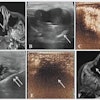
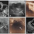
Vascular Diagnosis with Ultrasound offers high quality, vivid images and anatomical figures in color, as well as concise reference tables. For example, Vmax, Vmean, RI, and maximum diameter of the major abdominal arteries (aorta, celiac, splenic common hepatic, left gastric arteries, etc.) are listed in one table, which makes for easy referencing.
AuntMinnie.com contributing writer
January 29, 2002
Dr H. Le is a radiologist in private practice in Calgary, Alberta, Canada.
If you are interested in reviewing a book, let us know at [email protected].
The opinions expressed in this review are those of the author, and do not necessarily reflect the views of AuntMinnie.com.
Copyright © 2002 AuntMinnie.com

