Semiinvasive pulmonary aspergillosis in chronic obstructive pulmonary disease: radiologic and pathologic findings in nine patients.
Franquet T, Muller NL, Gimenez A, Domingo P, Plaza V, Bordes R
OBJECTIVE: The purpose of this study is to assess the radiographic,
thin-section CT, and histologic findings of semiinvasive aspergillosis
in patients with chronic obstructive pulmonary disease (COPD). MATERIALS
AND METHODS: The study included nine patients with COPD seen at the Hospital
de Sant Pau during a 3-year period who had histopathologically proven aspergillosis
with tissue invasion. Chest radiography and thin-section (2-mm collimation)
CT of the chest were available in all cases. RESULTS: Nine patients had
semiinvasive aspergillosis proven at autopsy (n = 7) or by thoracoscopically
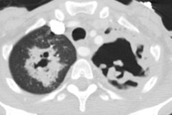
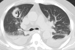
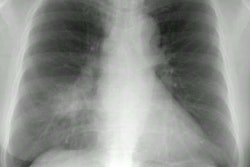
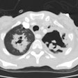
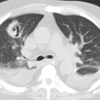
guided lung biopsy (n = 2). The radiologic findings consisted of parenchymal
consolidation (n = 6) and nodules larger than 1 cm in diameter (n = 3).
Parenchymal consolidation involved the upper lobes in five patients and
was bilateral in four. Cavitation was present in two of the patients with
consolidation and in two of the patients with nodular opacities. Adjacent
pleural thickening was revealed by CT in four patients. Histologically,
the areas of consolidation represented active inflammation and intraalveolar
hemorrhage containing Aspergillus organisms. In the three patients with
multiple cavitated nodules, a variable degree of central necrosis was observed.
The inflammatory infiltrate extended into the surrounding lung parenchyma,
and adjacent areas of hemorrhage were also seen. Aspergillus colonies were
identified within the lung tissue. CONCLUSION: Upper lobe consolidation
or multiple nodules in patients with COPD should raise the possibility
of semiinvasive aspergillosis.
