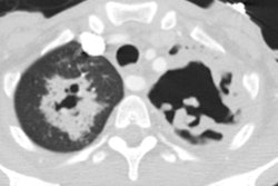
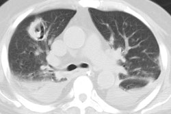
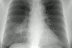
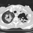
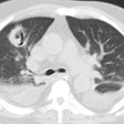
The radiographic findings of fibrosing mediastinitis.
Sherrick AD, Brown LR, Harms GF, Myers JL
We retrospectively reviewed the radiographic findings of fibrosing mediastinitis (FM) in 33 patients. Imaging studies included chest radiographs, computed tomographic scans, magnetic resonance imaging examinations, esophograms, ventilation perfusion scans, angiograms, and venograms. Findings include bronchial narrowing in 11 patients (33 percent), pulmonary artery obstruction/narrowing in 6 patients (18 percent), esophageal narrowing in 3 patients (9 percent), and superior vena cava obstruction/narrowing in 13 patients (39 percent). Two distinctly different radiographic patterns were identified: a localized pattern seen in 27 patients (82 percent) that frequently contained calcification and a diffuse pattern seen in 6 patients (18 percent) that did not contain calcification. The localized pattern is most likely due to histoplasmosis and does not show radiographic evidence of improvement with steroid therapy. The diffuse pattern may more likely be truly idiopathic or of a noninfectious etiology. Several patients with the diffuse pattern showed radiographic evidence of improvement with steroid therapy.
