Radiology 1995 Aug;196(2):409-414. Invasive pulmonary aspergillosis in AIDS: radiographic, CT, and pathologic findings.
Staples CA, Kang EY, Wright JL, Phillips P, Muller NL
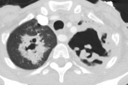
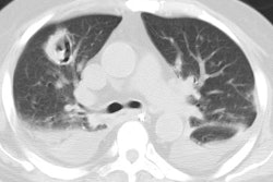
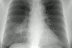
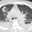
PURPOSE: To review the radiographic and computed tomographic (CT) manifestations of invasive pulmonary aspergillosis and to correlate the imaging and pathologic findings in patients with acquired immunodeficiency syndrome (AIDS). MATERIALS AND METHODS: Chest radiographs, CT scans, and pathologic specimens were reviewed retrospectively in 10 AIDS patients with proved invasive pulmonary aspergillosis. RESULTS: The most common radiographic finding was the presence of thick-walled cavitary lesions. Less common findings included nodules, consolidation, and pleural effusion. CT depicted more nodules and cavities than did radiography. The predominant pathologic abnormalities consisted of tissue invasion and abscess formation and angioinvasion with or without infarction. All patients had infection with Aspergillus fumigatus as well as other pathogens, the most common being cytomegalovirus and Pseudomonas aeruginosa. CONCLUSION: Thick-walled cavitary lesions are the most common radiologic manifestation of invasive pulmonary aspergillosis in AIDS. The findings are more numerous and better defined on CT scans. The radiologic findings reflect a spectrum of pathologic abnormalities.
PMID: 7617853, MUID: 95343104

