Tracheobronchopathia Osteochondroplastica:
View cases
Clinical:
This is a rare (0.02 to 0.7% of patients that undergo bronchoscopy [3]), non-neoplastic disorder that is characterized by the presence of small 2 to 5 mm osteocartilagenous nodules within the submucosa of the lower two-thirds of the trachea, and to a lesser degree the proximal bronchi. Serum calcium and phosphate levels are normal. Men are affected more than women (3:1) and over 50% of affected patients are more than 50 years old. Only about 10% of affected patients are symptomatic and present with dyspnea, cough, recurrent pneumonia, or hemoptysis. The disease is quite indolent and is a benign condition with no reported malignant transformation. The nodules are enchondroses that arise from the tracheobronchial cartilagenous rings and are continuous with the perichondrium of the underlying cartilage (posterior membranous trachea is not affected by this disorder). The nodules are composed of hyaline cartilage, areas of lamellar bone, and sometimes contain hematopoietic marrow. Treatment is silicone rubber prosthetic stents as dilatation alone results in rapid recurrence.
Tracheobronchopathia osteochondroplastica should not be confused with the normal tracheal and bronchial calcification that can be seen in patients over the age of 40 years. There is an increased incidence of tracheal calcification in patients receiving warfarin therapy and tracheal calcification can also occur in children and young adults on long term warfarin [5].
X-ray:
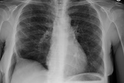
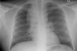
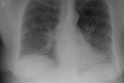
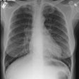
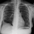
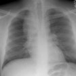
Chest radiographs are typically normal [4], but may reveal narrowing of the involved airway and may demonstrate the presence of calcifications within the airway. CT will demonstrate the small, sessile calcified intralumenal masses arising from the anterior and lateral walls, with sparing of the posterior membranous tracheal wall (unlike amyloid involvement) [4].
REFERENCES:
(1) J Thorac Imaging 1995;10(3):180-198 (p.194-196)
(2) J Thorac Imaging 1995;10(4):236-254 (p.238)
(3) Radiographics 2002; Nonneoplastic lesions of the tracheobronchial wall: radiologic findings with bronchoscopic correlation. 22: S215-S230
(4) Radiographics 1998; Meyer CA, White CS. Cartilagenous disorders of the chest. 18: 1109-1123
(5) AJR 2000; Joshi A, et al. CT detection of tracheobronchial calcification in an 18-year-old on maintenence warfarin sodium therapy: Cause and effect? 175: 921

