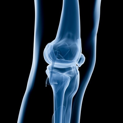
RSIP Vision is highlighting preliminary results from an ongoing clinical study evaluating the potential of an artificial intelligence (AI) tool to reconstruct a 3D knee model from 2D x-ray images at Assuta Medical Center in Tel Aviv.
This tool is based on novel, patent-pending technology that uses AI to produce a model of the knee from two x-ray images. The company said this model can be used for surgical planning and navigation during orthopedic procedures, such as total knee replacement.
RSIP plans to expand the clinical study to a U.S. medical center and hopes to eventually receive clearance from the U.S. Food and Drug Administration. It is also working to extend this software to additional anatomical regions, such as the hip and shoulder joints.







![A normal mammogram confirmed by three-year radiologic follow-up illustrates reader-marked regions of interest (ROIs) during (A) unaided (round 1) and (B) artificial intelligence (AI)–assisted (round 2) reading. Each colored dot represents an ROI for recall by a human reader. Readers could mark more than one ROI per case, represented by multiple dots of the same color. During AI-assisted reading, the AI system displayed three visible prompts: two with suspicion of malignancy scores of 35% (left mediolateral oblique [L MLO] and craniocaudal [L CC]) and one with a suspicion of malignancy score of 10% (right craniocaudal [R CC]), shown as polygonal overlays. Without AI, six of 10 readers (60%) marked a false-positive ROI. With AI assistance, this fell to two of 10 (20%). R MLO = right mediolateral oblique.](https://img.auntminnie.com/mindful/smg/workspaces/default/uploads/2026/07/2026-07-14-radiology-mammogram-ai-auto-bias.H0bYO8QlWs.jpg?auto=format%2Ccompress&fit=crop&h=112&q=70&w=112)


