Dear AuntMinnie Member,
Extreme physical conditioning of the type employed by triathletes may produce distinct changes in cardiac anatomy that can be detected on MRI scans, according to a new study we're featuring this week in our Cardiac Imaging Digital Community.
German researchers used 1.5-tesla MRI to examine triathletes and found that their hearts developed greater muscle mass and thickness, as well as other changes. The changes are among the first to be detected by cardiac MRI, according to the research group.
Learn why these findings are important by clicking here, or visit the Cardiac Imaging Digital Community at cardiac.auntminnie.com.
CAD for abdominal CT
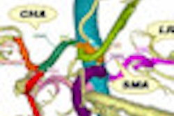
In other news, in our Advanced Visualization Digital Community we're reporting on a new computer-aided detection (CAD) method for segmenting and labeling abdominal arteries.
Japanese researchers developed the algorithm to save time, reduce errors, and improve clinicians' understanding of the complex anatomy and branching patterns of the abdominal arteries. They believe it's an improvement over previous CAD schemes that were based on position information of organs.
Learn more by clicking here, or visit the Advanced Visualization Digital Community at av.auntminnie.com.



![A normal mammogram confirmed by three-year radiologic follow-up illustrates reader-marked regions of interest (ROIs) during (A) unaided (round 1) and (B) artificial intelligence (AI)–assisted (round 2) reading. Each colored dot represents an ROI for recall by a human reader. Readers could mark more than one ROI per case, represented by multiple dots of the same color. During AI-assisted reading, the AI system displayed three visible prompts: two with suspicion of malignancy scores of 35% (left mediolateral oblique [L MLO] and craniocaudal [L CC]) and one with a suspicion of malignancy score of 10% (right craniocaudal [R CC]), shown as polygonal overlays. Without AI, six of 10 readers (60%) marked a false-positive ROI. With AI assistance, this fell to two of 10 (20%). R MLO = right mediolateral oblique.](https://img.auntminnie.com/mindful/smg/workspaces/default/uploads/2026/07/2026-07-14-radiology-mammogram-ai-auto-bias.H0bYO8QlWs.jpg?auto=format%2Ccompress&fit=crop&h=100&q=70&w=100)


![A normal mammogram confirmed by three-year radiologic follow-up illustrates reader-marked regions of interest (ROIs) during (A) unaided (round 1) and (B) artificial intelligence (AI)–assisted (round 2) reading. Each colored dot represents an ROI for recall by a human reader. Readers could mark more than one ROI per case, represented by multiple dots of the same color. During AI-assisted reading, the AI system displayed three visible prompts: two with suspicion of malignancy scores of 35% (left mediolateral oblique [L MLO] and craniocaudal [L CC]) and one with a suspicion of malignancy score of 10% (right craniocaudal [R CC]), shown as polygonal overlays. Without AI, six of 10 readers (60%) marked a false-positive ROI. With AI assistance, this fell to two of 10 (20%). R MLO = right mediolateral oblique.](https://img.auntminnie.com/mindful/smg/workspaces/default/uploads/2026/07/2026-07-14-radiology-mammogram-ai-auto-bias.H0bYO8QlWs.jpg?auto=format%2Ccompress&fit=crop&h=112&q=70&w=112)