Dear AuntMinnie Member,
Yet another clinical study has confirmed the safety of contrast media for echocardiography studies -- the question is whether the U.S. Food and Drug Administration (FDA) is listening.
The FDA in 2007 required that echocardiography contrast products carry black box warnings after four deaths were reported in patients who received contrast media. However, several studies published since then have failed to find a connection between patient mortality and echo contrast.
In the latest research, a team from Michigan performed a meta-analysis of eight studies that covered more than 5 million patients, and like some of the other studies, the group actually found lower mortality rates in the patients who received contrast compared to those who didn't.
Will the results be enough to persuade the FDA to remove its black box warning? We don't know about that, but we can point you to the article by clicking here, or visit our Cardiac Imaging Digital Community at cardiac.auntminnie.com.
CAD for virtual colonoscopy
One area where the FDA has started to clear a regulatory logjam is in computer-aided detection (CAD) software. The agency has begun issuing clearances of CAD products after several years of inactivity, giving heart to proponents of this technology.
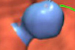
One such recently cleared product is a CAD application for virtual colonoscopy studies, and we're featuring an article about the software in our Virtual Colonoscopy Digital Community. Researchers from the University of Chicago put the software through its paces in a recent clinical study, finding that most of their 19 readers reported higher sensitivity with the algorithm than without it.
Read all about it by clicking here, or visit the community at vc.auntminnie.com.

![A normal mammogram confirmed by three-year radiologic follow-up illustrates reader-marked regions of interest (ROIs) during (A) unaided (round 1) and (B) artificial intelligence (AI)–assisted (round 2) reading. Each colored dot represents an ROI for recall by a human reader. Readers could mark more than one ROI per case, represented by multiple dots of the same color. During AI-assisted reading, the AI system displayed three visible prompts: two with suspicion of malignancy scores of 35% (left mediolateral oblique [L MLO] and craniocaudal [L CC]) and one with a suspicion of malignancy score of 10% (right craniocaudal [R CC]), shown as polygonal overlays. Without AI, six of 10 readers (60%) marked a false-positive ROI. With AI assistance, this fell to two of 10 (20%). R MLO = right mediolateral oblique.](https://img.auntminnie.com/mindful/smg/workspaces/default/uploads/2026/07/2026-07-14-radiology-mammogram-ai-auto-bias.H0bYO8QlWs.jpg?auto=format%2Ccompress&fit=crop&h=100&q=70&w=100)


![A normal mammogram confirmed by three-year radiologic follow-up illustrates reader-marked regions of interest (ROIs) during (A) unaided (round 1) and (B) artificial intelligence (AI)–assisted (round 2) reading. Each colored dot represents an ROI for recall by a human reader. Readers could mark more than one ROI per case, represented by multiple dots of the same color. During AI-assisted reading, the AI system displayed three visible prompts: two with suspicion of malignancy scores of 35% (left mediolateral oblique [L MLO] and craniocaudal [L CC]) and one with a suspicion of malignancy score of 10% (right craniocaudal [R CC]), shown as polygonal overlays. Without AI, six of 10 readers (60%) marked a false-positive ROI. With AI assistance, this fell to two of 10 (20%). R MLO = right mediolateral oblique.](https://img.auntminnie.com/mindful/smg/workspaces/default/uploads/2026/07/2026-07-14-radiology-mammogram-ai-auto-bias.H0bYO8QlWs.jpg?auto=format%2Ccompress&fit=crop&h=112&q=70&w=112)