With the advantages of portable radiography, namely the ability to transport the x-ray system to the patient in a wide range of situations, come inherent disadvantages.
In order to minimize scatter, portable x-ray system operators must be well-versed in imaging geometry and anatomy to effectively apply complex antiscatter grids to accommodate a range of patients and situations.
Researchers at Emory University's Winship Cancer Institute in Atlanta and GE Global Research of Niskayuna, NY, are collaborating on a software-based method for reducing scatter in portable systems. Tested on thorax phantom images, the algorithm reduced the x-ray signal by 50% to 100% without loss of image quality, according to Ioannis Sechopoulos, Ph.D., assistant professor of radiology at Emory University School of Medicine, who presented the study at the recent American Association of Physicists in Medicine (AAPM) meeting in Houston.
When the method was applied to images of a thorax phantom obtained with a portable x-ray system without a grid, image quality was comparable to the quality obtained with a fixed clinical system with a grid, said Sechopoulos.
 |
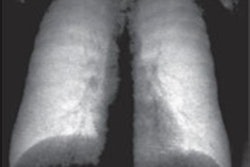
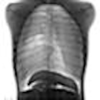
| Chest anteroposterior images of a thorax phantom acquired (a) with a clinical system with an antiscatter grid and (b) with a portable system without an antiscatter grid; (c) image in (b) after processing with the proposed method. Images provided by Ioannis Sechopoulos, Ph.D., Emory University School of Medicine, Winship Cancer Institute. |
After studying x-ray scatter signals present in different portable radiography systems, the researchers confirmed that the x-ray scatter fields consisted of a low-frequency offset, with various shapes and magnitudes depending on the image being acquired.
The algorithm developed by the researchers is a modified version of unsharp masking that involves masking the open field area and the thin sections of the body, and replacing these areas with the mean signal from the edges of the body. Masking prevents the very high signal areas from being included in the low pass image, which would lead to a false estimation of the signal to be removed. Compensation is given for the addition of a low pass filter.
The researchers tested the algorithm's effectiveness by comparing the difference between the original image and the processed image with the results of repeated random sampling of the scatter-only image. They also computed the mean reduction in different regions-of-interest (ROI) images.
"The developed scatter reduction algorithm seems to compensate for the inability to use a high ratio grid in portable systems," said Sechopoulos. The researchers plan future testing on chest and other phantoms before advancing to human testing.
By Kathlyn Stone
AuntMinnie.com contributing writer
August 22, 2008
Related Reading
Dark-field x-ray measures scatter radiation for better image quality, February 4, 2008
CR/DR image quality: Issues and concerns, April 12, 2007
Antiscatter grids may not be helpful in some DR applications, September 22, 2006
Copyright © 2008 AuntMinnie.com