Dear Digital X-Ray Insider,
In this edition of the Insider, we're offering a profile of a company with a somewhat unique sales proposition: that its video fluoroscopy system can produce higher legal settlements for patients suffering from whiplash.
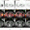
The company, DMX Works of Palm Harbor, FL, claims that its Digital Motion X-ray system reveals details of spinal motion that can detect underlying sources of neck pain not visualized with CT or MRI. But is the system really the best clinical choice for this application? And how much radiation dose does the system produce? Find how by clicking here for an Insider Exclusive by associate editor Donna Domino.
In other news in the community this month, learn about a recent clinical study on the use of computer-aided detection (CAD) software for chest computed radiography (CR) exams. Researchers found that adding CAD as a second reader improved radiologist accuracy, particularly specificity. Learn more by clicking here.
Other recent articles in the community include:
- A story on the value of radiography in diagnosing the severity of knee osteoarthritis
- The contribution of a pair of teens to the field of radiology research
- A story on the superiority of CR-based mammography over analog for screening applications
Get these stories and more in your Digital X-Ray Community at xray.auntminnie.com.