Dear X-Ray Insider,
When the subject is a complex and moving target -- such as whether to convert to digital x-ray via CR or DR -- it always makes for interesting reading.
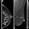
Researchers have now published the most ambitious comparison of CR and DR to date, looking at several different systems in varying settings. That alone made it a worthwhile choice for this month's X-Ray Insider Exclusive, delivered to you before it reaches other AuntMinnie.com members.
But what makes this report even more interesting is the detail it offers on how some facilities take up to six minutes longer than others to generate a simple two-view chest study.
Perhaps the wasted time isn't an issue. After all, the authors note that all of the facilities they examined -- and many others -- are operating at far less than capacity.
On the other hand, general radiography still comprises the bulk of imaging volume, according to the authors, and yet reportedly costs more to perform than it garners in Medicare reimbursement. You would think those economic pressures, along with a growing shortage of x-ray technologists, would eventually inspire greater interest in operational efficiency.
If digital conversion or departmental efficiency is on your agenda, you'll want to check out this story in the X-Ray Digital Community by clicking here. And as always, I welcome your feedback and story suggestions.
![Images show the pectoralis muscles of a healthy male individual who never smoked (age, 66 years; height, 178 cm; body mass index [BMI, calculated as weight in kilograms divided by height in meters squared], 28.4; number of cigarette pack-years, 0; forced expiratory volume in 1 second [FEV1], 97.6% predicted; FEV1: forced vital capacity [FVC] ratio, 0.71; pectoralis muscle area [PMA], 59.4 cm2; pectoralis muscle volume [PMV], 764 cm3) and a male individual with a smoking history and chronic obstructive pulmonary disorder (COPD) (age, 66 years; height, 178 cm; BMI, 27.5; number of cigarette pack-years, 43.2, FEV1, 48% predicted; FEV1:FVC, 0.56; PMA, 35 cm2; PMV, 480.8 cm3) from the Canadian Cohort Obstructive Lung Disease (i.e., CanCOLD) study. The CT image is shown in the axial plane. The PMV is automatically extracted using the developed deep learning model and overlayed onto the lungs for visual clarity.](https://img.auntminnie.com/mindful/smg/workspaces/default/uploads/2026/03/genkin.25LqljVF0y.jpg?auto=format%2Ccompress&crop=focalpoint&fit=crop&h=100&q=70&w=100)


![Images show the pectoralis muscles of a healthy male individual who never smoked (age, 66 years; height, 178 cm; body mass index [BMI, calculated as weight in kilograms divided by height in meters squared], 28.4; number of cigarette pack-years, 0; forced expiratory volume in 1 second [FEV1], 97.6% predicted; FEV1: forced vital capacity [FVC] ratio, 0.71; pectoralis muscle area [PMA], 59.4 cm2; pectoralis muscle volume [PMV], 764 cm3) and a male individual with a smoking history and chronic obstructive pulmonary disorder (COPD) (age, 66 years; height, 178 cm; BMI, 27.5; number of cigarette pack-years, 43.2, FEV1, 48% predicted; FEV1:FVC, 0.56; PMA, 35 cm2; PMV, 480.8 cm3) from the Canadian Cohort Obstructive Lung Disease (i.e., CanCOLD) study. The CT image is shown in the axial plane. The PMV is automatically extracted using the developed deep learning model and overlayed onto the lungs for visual clarity.](https://img.auntminnie.com/mindful/smg/workspaces/default/uploads/2026/03/genkin.25LqljVF0y.jpg?auto=format%2Ccompress&crop=focalpoint&fit=crop&h=112&q=70&w=112)


