Elsevier Science, St. Louis, 2002, $39.95
Case Review: Gastrointestinal Imaging by Peter J. Feczko and Robert D. Halpert
Elsevier Science, St. Louis, 2000, $39.95
The latest installments in the Case Review Series adhere to the master design of the series. Two cases are presented on one page, each consisting of one or two images and four related questions. This is followed by brief answers, a case commentary, references to relevant articles from the radiology literature (often review articles), and promotional cross-references to the popular Requisites series.
The cases are presented by level of difficulty. Graduating radiology residents should easily solve the "Opening Round" cases. "Fair Game" cases are a more complicated but still solvable mix. The final section of "Challenge" cases is designed to test fellows and senior residents on their powers of differential diagnosis. Indices of cases and terminologies round out each volume. The design lends itself equally well to preparation for the boards or as refreshers. It’s possible to review a case, a section or even an entire volume in a single sitting.
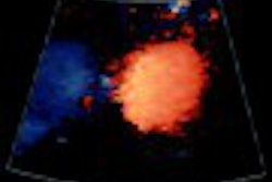
General and Vascular Ultrasound is a remarkable and welcome addition to the library of review in sonography. Most wonderful is the uncommonly beautiful image quality throughout -- Dr. Middleton personally scanned each case and he gave the images the same attention that he lavished on the text.
Case questions are written to draw attention to image findings, but without divulging the diagnosis prematurely. More than two hundred cases include a survey of normal and pathologic anatomy of the abdomen, retroperitoneum, vascular tree and small parts including prostate, testes, and thyroid (obstetric and gynecologic ultrasound are reviewed in a separate volume by different authors).
The numerous musculoskeletal cases are a boon to those who are unfamiliar with sonographic applications in this body system. Normal anatomy of the shoulder is emphasized along with entities such as rotator cuff tears, biceps tendon rupture, Morrant-Baker cysts, Achilles tendon tears, and plantar fasciitis.
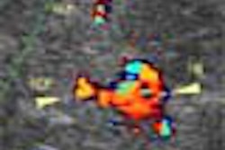
A number of cases focus on the recognition of common artifacts and technical errors encountered in gray scale and color Doppler images. These cases guide the reader to a clearer understanding of the technical factors important in producing high quality, diagnostic ultrasound images.
One drawback of the book is that all the color Doppler plates are collated in an appendix. The author bemoaned this fact in his preface, and I found it inconvenient as the reader must flip from the gray scale image printed in the case to the referenced color images located in a different part of the book. Still, General and Vascular Ultrasound is a real bang for the buck, clearly surpassing its objective of mere review and providing a high quality teaching file on sonography.
Drs. Feczko and Halpert have undertaken an enormous task with Gastrointestinal Imaging, selecting two hundred cases for review from this broad topic. The results are admirable.
At its core is a wide selection of barium cases illustrating pathology from hypopharynx to anus, with ample attention given to the dreaded differential diagnosis of small bowel abnormalities. CT cases round out the bulk of the work, with a smattering of classic radiographic diagnoses and a few examples using sonography, MRI, nuclear medicine and visceral angiography.
Image quality is generally good, but in some cases the findings are difficult to see due to the small size of the abdominal radiograph reproduction, or the small and disorienting field-of-view in coned-down images.
Case questions often hint at the diagnosis but do not always specifically focus the reader on the abnormal findings depicted on the images; these findings are often likewise omitted from the written case discussions, which focus on the disease processes instead. These are minor shortcomings of the volume, however, since its clear articulation of abnormal findings forms the basis for subsequent accurate interpretation.
Case commentaries are well written and informative, and a single literature reference is cited for each. Several examples of defecography studies should be of special interest to readers who have neither seen nor performed this type of study. However, a case illustrating normal defecographic anatomy, and discussing performance of the exam would have been useful.
The authors’ method of posing questions is clever, provocative, and instructive. For example, regarding free intraperitoneal fluid, they ask: "What renal abnormality could cause this abnormality?" Answer: "None, kidneys are retroperitoneal." Overall, Gastrointestinal Imaging is well worth the reader’s investment in time and money.
Dr. Raymond ThorntonAuntMinnie.com contributing writer
October 17, 2002
Dr. Thornton, a graduate of the University of Pittsburgh School of Medicine, is currently chief resident, department of radiology, at the University of California, San Francisco. He also will be a fellow in vascular and interventional radiology at UCSF.
If you are interested in reviewing a book, let us know at [email protected].
The opinions expressed in this review are those of the author, and do not necessarily reflect the views of AuntMinnie.com.
Copyright © 2002 AuntMinnie.com