(Radiology Review) Korean radiologists have described the ultrasound findings of pelvic congestion syndrome (PCS) in a prospective study published in the American Journal of Roentgenology.
Ten percent of patients presenting to gynecologists complain of pelvic pain, and pelvic varicoceles are found in half of the women with pelvic pain, according to Dr. Song Jin Park and colleagues.
Thirty-two patients with pelvic congestion syndrome and 35 age-matched healthy volunteers were evaluated with transabdominal and transvaginal ultrasound, which was performed at least one month before selective ovarian venography. Clinical features and findings on selective ovarian venography were used to determine PCS.
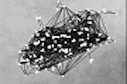
Features assessed included "ovarian vein diameter and flow direction, presence of pelvic varicocele, diameter of the pelvic veins, change of the duplex waveform during the Valsalva's maneuver, volume of the uterus, and presence of polycystic changes in the ovaries." The authors successfully demonstrated the left ovarian vein draining into the left renal vein in 31 of 32 women with PCS.
"On transvaginal sonography, pelvic varicoceles were present in all patients with pelvic congestion syndrome (100%) and in six control subjects (17.1%)," they said.
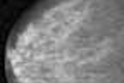
The authors considered an ovarian vein diameter of 5 mm the upper limit of normal. They found large veins crossing the uterine myometrium in eight patients with pelvic congestion syndrome (25%). Three control subjects also had large veins crossing the uterine myometrium.
"Communication between bilateral pelvic varicosities via transuterine crossing veins is a common selective ovarian venography finding in pelvic congestion syndrome," they reported, and "the presence of crossing veins on transvaginal sonography correlates with large crossing veins that permit contralateral filling of contrast media on selective ovarian venography."
Compared with the control group, women with PCS had three times the rate of polycystic ovarian change. About half of the women with PCS had cystic ovarian changes but there was no significant difference in uterine volume comparing the two groups.
According to the authors, "sonographic findings of pelvic congestion syndrome were dilated left ovarian vein with reversed caudal flow, presence of varicocele, dilated arcuate veins crossing the uterine myometrium, polycystic changes of the ovary, and variable duplex waveform during the Valsalva's maneuver."
A combination of transabdominal and transvaginal ultrasound imaging provides a noninvasive selection method. Patients suffering chronic pelvic pain may proceed to ovarian venography and transcatheter embolization, depending on the sonographic findings, they suggested.
Diagnosis of pelvic congestion syndrome using transabdominal and transvaginal sonographySeong Jin Park, et. al.
Department of radiology, Soonchunhyang University Bucheon Hospital, Gyeonggi-do, Republic of Korea.
AJR 2004 March; 182:683-688
By Radiology Review
March 14, 2004
Copyright © 2004 AuntMinnie.com