CHICAGO - Gastroenterologists said it couldn't be done. Or that it might be possible, but no one really knew. So radiologists attending Monday's RSNA virtual colonoscopy session were delighted to hear that VC detected superficial (nonpolypoid) colonic lesions as well as conventional colonoscopy.
The first study designed specifically to gauge CT's ability to detect the hard-to-find lesions was led by Dr. Nobuyuki Shiraga, chief of radiology at Tachikawa Kyosai Hospital in Tokyo. "The purpose of this presentation is to assess the detectability of superficial lesions in CT colonography using multidetector-row CT datasets," he said.
From July 2000 to August 2002, 179 subjects with suspected colonic lesions underwent virtual colonoscopy followed by same-day conventional colonoscopy. The patients had undergone standard bowel preparation.
The group used a LightSpeed QX/i 16-slice multislice scanner (GE Medical Systems, Waukesha, WI), using 2.5-mm collimation and a pitch of 3.
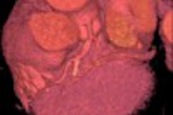
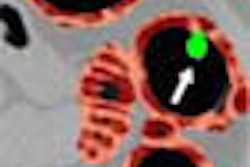
"A total of 37 superficial lesions were depicted," Shiraga said. "There were nine cases of colorectal carcinoma, and they were all depicted with CT colonoscopy."
According to an abstract accompanying the presentation, other lesions detected in virtual colonoscopy included a tubular adenoma with severe dysplasia, four tubular adenomas with moderate dysplasia, and nine cases of hyperplastic colonic epithelium. One mucosal-stage cancer was not depicted with VC even in a retrospective assessment, according to the abstract.
Five lesions were not seen in virtual colonoscopy, Shiraga said. These included two cases of tubular adenoma smaller than 9 mm, one case of hyperplastic colonic epithelium smaller than 9 mm, and two cases of hyperplastic epithelium larger than 10 mm.
"Multidetector CT colonography has a high potential to detect superficial colonic lesions including de novo cancer," Shiraga concluded.
"You confirmed that a significant number of (superficial) lesions are in fact hyperplastic, and that's very interesting," commented Dr. Abraham Dachman from the University of Chicago. "Looking at your images confirms my suspicion that we need to come up with a new classification of flat lesions, because there is only one definition in the literature, and clearly some flat lesions are polypoid and some are not." The problem is that sensitivity figures will vary depending on how these lesions are classified, Dachman said.
As for specificity, Shiraga said it could not be reliably measured in the current study.
By Eric Barnes
AuntMinnie.com staff writer
December 3, 2002
Copyright © 2002 AuntMinnie.com